. 2023 Oct;299(10):105209.
doi: 10.1016/j.jbc.2023.105209. Epub 2023 Sep 1.

The biased M3 mAChR ligand PD 102807 mediates qualitatively distinct signaling to regulate airway smooth muscle phenotype

Affiliations

The biased M3 mAChR ligand PD 102807 mediates qualitatively distinct signaling to regulate airway smooth muscle phenotype

Eric Tompkins et al. J Biol Chem. 2023 Oct.

Abstract

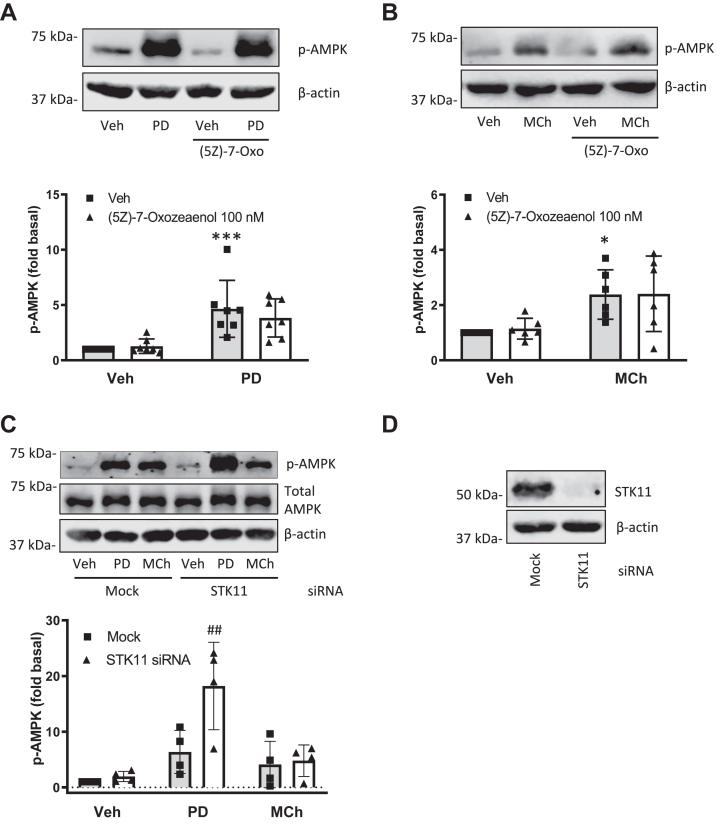
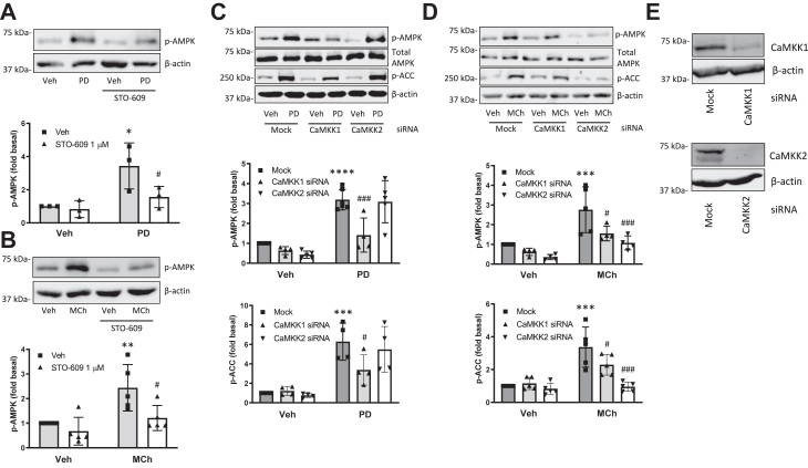
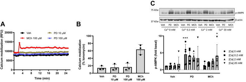
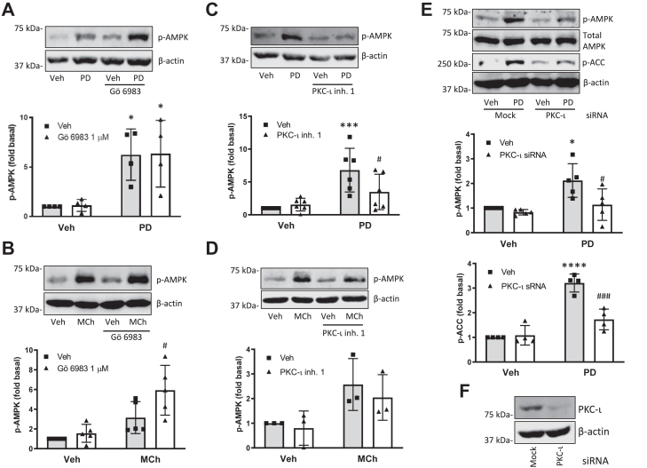
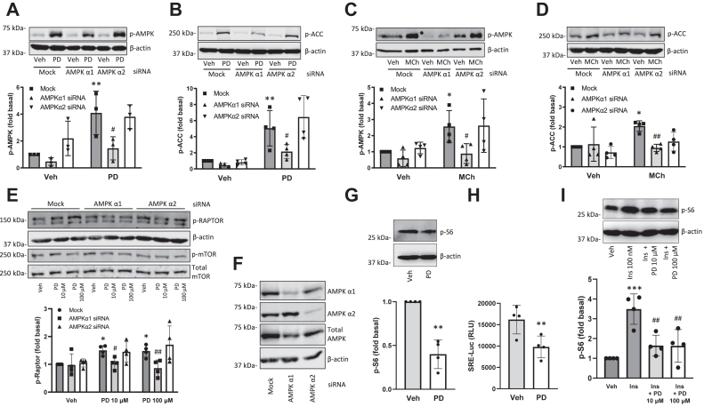
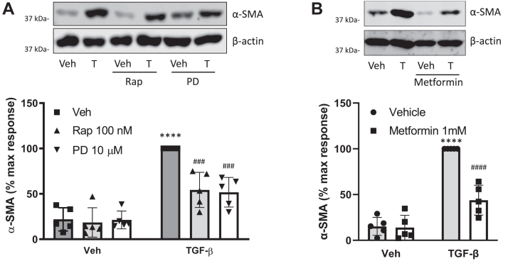
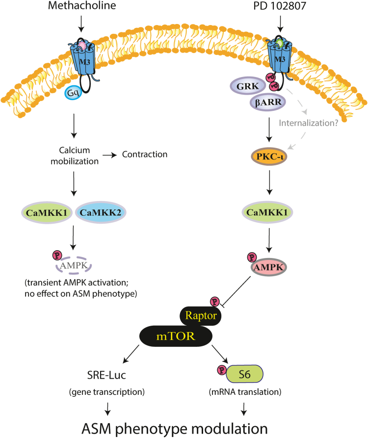
Airway smooth muscle (ASM) cells attain a hypercontractile phenotype during obstructive airway diseases. We recently identified a biased M3 muscarinic acetylcholine receptor (mAChR) ligand, PD 102807, that induces GRK-/arrestin-dependent AMP-activated protein kinase (AMPK) activation to inhibit transforming growth factor-β-induced hypercontractile ASM phenotype. Conversely, the balanced mAChR agonist, methacholine (MCh), activates AMPK yet does not regulate ASM phenotype. In the current study, we demonstrate that PD 102807- and MCh-induced AMPK activation both depend on Ca2+/calmodulin-dependent kinase kinases (CaMKKs). However, MCh-induced AMPK activation is calcium-dependent and mediated by CaMKK1 and CaMKK2 isoforms. In contrast, PD 102807-induced signaling is calcium-independent and mediated by the atypical subtype protein kinase C-iota and the CaMKK1 (but not CaMKK2) isoform. Both MCh- and PD 102807-induced AMPK activation involve the AMPK α1 isoform. PD 102807-induced AMPK α1 (but not AMPK α2) isoform activation mediates inhibition of the mammalian target of rapamycin complex 1 (mTORC1) in ASM cells, as demonstrated by increased Raptor (regulatory-associated protein of mTOR) phosphorylation as well as inhibition of phospho-S6 protein and serum response element-luciferase activity. The mTORC1 inhibitor rapamycin and the AMPK activator metformin both mimic the ability of PD 102807 to attenuate transforming growth factor-β-induced α-smooth muscle actin expression (a marker of hypercontractile ASM). These data indicate that PD 102807 transduces a signaling pathway (AMPK-mediated mTORC1 inhibition) qualitatively distinct from canonical M3 mAChR signaling to prevent pathogenic remodeling of ASM, thus demonstrating PD 102807 is a biased M3 mAChR ligand with therapeutic potential for the management of obstructive airway disease.

Keywords: G protein-coupled receptor; airway; airway smooth muscle; asthma; biased receptor pharmacology.


Conflict of interest statement

Conflict of interest The authors declare that they have no conflicts of interest with the contents of this article.

Figures

Figure 1
Figure 1
AMPK kinases TAK1 and STK11 (LKB1) do not mediate M3 mAChR-induced AMPK phosphorylation.A and B, cells were preincubated with 5z-7-oxozeaenol (100 nM) for 15 min then stimulated with (A) PD 102807 (100 μM; t = 20 min) or (B) MCh (100 μM; t = 5 min). C, following siRNA transfection against STK11 (as described in Experimental procedures), cells were stimulated with PD 102807 or MCh. D, STK11 knockdown was confirmed by immunoblotting. Representative blots are shown. Loading was corrected for β-actin or total AMPK. Data are means ± SD. ∗p < 0.05, ∗∗∗ p < 0.001 versus vehicle stimulation; ##p < 0.01 versus mock-transfected PD 102807; one-way ANOVA followed by Bonferroni multiple comparison test. AMPK, AMP-activated protein kinase; LKB1, liver kinase B1; mAChR, muscarinic acetylcholine receptor; MCh, methacholine.
Figure 2
Figure 2
CaMKKs mediate M3 mAChR-induced AMPK phosphorylation.A and B, cells were preincubated with STO-609 (1 μM) for 15 min and then stimulated with (A) PD 102807 (100 μM; t = 20 min) or (B) MCh (100 μM; t = 5 min). C and D, cells were transfected with siRNA against CaMKK1 or CaMKK2 and then stimulated with (C) PD 102807 or (D) MCh. AMPK and ACC phosphorylation was determined using phospho-specific antibodies. E, knockdown of CaMKK isoforms was confirmed by immunoblotting. Representative blots are shown. Loading was corrected for total AMPK or β-actin. Data are means ± SD. ∗p < 0.05, ∗∗p < 0.01, ∗∗∗ p < 0.001, ∗∗∗∗p < 0.0001 versus vehicle stimulation, #p < 0.05, ###p < 0.001 versus respective mock-transfected PD 102807 or MCh; one-way ANOVA followed by Bonferroni multiple comparison test. ACC, acetyl-CoA carboxylase; AMPK, AMP-activated protein kinase; CaMKKs, Ca2+/calmodulin-dependent kinase kinases; mAChR, muscarinic acetylcholine receptor; MCh, methacholine.
Figure 3
Figure 3
PD 102807-induced p-AMPK is independent of calcium mobilization.A and B, cells were stimulated with PD 102807 (10 and 100 μM) or MCh (100 μM), and calcium mobilization was measured using fluo-4 AM calcium indicator and Flexstation III as described in Experimental procedures. C, cells were permeabilized using β-escin (100 nM) then treated with increasing concentrations of CaCl2 (0, 0.2, 2, and 20 mM) and allowed to equilibrate for 5 min. Cells were then stimulated with PD 102807 (100 μM; t = 20 min) or MCh (100 μM; t = 5 min). Cells were lysed, and phosphorylation status of AMPK was determined by immunoblotting. Representative blots are shown. Loading was corrected for β-actin. Data are means ± SD. ∗p < 0.05, ∗∗∗p < 0.001 versus vehicle stimulation; one-way ANOVA followed by Bonferroni multiple comparison test. AMPK, AMP-activated protein kinase; MCh, methacholine; p-AMPK, phospho-AMPK.
Figure 4
Figure 4
PD 102807 induces p-AMPK in a PKC-iota-dependent manner.A and B, cells were stimulated with (A) PD 102807 (100 μM; t = 20 min) or (B) MCh (100 μM; t = 5 min) in the presence or absence of the pan-PKC inhibitor Gö 6983 (1 μM; 15 min preincubation). C and D, cells were stimulated with (C) PD 102807 or (D) MCh in the presence or absence of the selective small molecule PKC-ι inhibitor 1 (10 μM; 15 min preincubation). Phosphorylation status of AMPK was determined using a phospho-specific AMPK antibody. E, cells were transfected with siRNA against PKC-ι and subsequently stimulated with PD 102807. Phosphorylation status of AMPK and ACC was determined using phospho-specific antibodies. F, PKC-ι knockdown was confirmed by immunoblotting. Representative blots are shown. Loading was corrected for β-actin or total AMPK. Data are means ± SD values from 3 to five experiments. ∗p < 0.05, ∗∗∗ p < 0.001, ∗∗∗∗p < 0.0001 versus vehicle stimulation, #p < 0.05, ###p < 0.001 versus respective mock-transfected PD 102807 or MCh; one-way ANOVA followed by Bonferroni multiple comparison test. ACC, ACC, acetyl-CoA carboxylase; AMPK, AMP-activated protein kinase; MCh, methacholine; p-AMPK, phospho-AMPK; PKC-ι, protein kinase C iota.
Figure 5
Figure 5
PD 102807 induces phosphorylation of AMPK alpha 1 and Raptor to inhibit mTOR signaling in ASM cells.AD, after siRNA transfection against AMPK α1 or α2 subunit cells were stimulated with (A and B) PD 102807 (100 μM; t = 20 min) or (C and D) MCh (100 μM; t = 5 min) and phosphorylation of (A and C) AMPK or (B and D) ACC was determined by immunoblotting using phospho-specific antibodies. E, after siRNA transfection against AMPK α1 or α2 subunit, cells were stimulated with PD 102807 (10 or 100 μM; t = 20 min) and expression of p-Raptor, β-actin, p-mTOR or total mTOR was determined by immunoblotting. F, AMPK α1 and α2 knockdown was confirmed by immunoblotting. G, PD 102807 (10 μM; 20 min stimulation) inhibits basal phosphorylation od S6 ribosomal protein. H, PD 102807 (10 μM; 18 h stimulation) inhibits basal SRE-Luc activity. I, PD 102807 (10 or 100 μM; 20 min pretreatment) inhibits insulin-induced (100 nM; 30 min) phosphorylation of S6 ribosomal protein. Representative blots are shown. Loading was corrected for β-actin or total AMPK. Data are means ± SD. ∗p < 0.05, ∗∗p < 0.01, ∗∗∗p < 0.001 versus vehicle stimulation, #p < 0.05, ##p < 0.01 versus respective vehicle pretreated, stimulated condition; one-way ANOVA followed by Bonferroni multiple comparison test; two-tailed Student’s t test. AMPK, AMP-activated protein kinase; ASM, Airway smooth muscle; MCh, methacholine; mTOR, the mammalian target of rapamycin; SRE-Luc, serum response element–driven luciferase activity.
Figure 6
Figure 6
mTORC1 inhibitor rapamycin and AMPK activator metformin inhibit TGF-β-induced alpha smooth muscleactinexpression.A and B, cells were stimulated for 3 days with TGF-β (1 ng/ml) in the presence or absence of (A) PD 102807 (10 μM) and rapamycin (100 nM) or (B) metformin (1 mM). Smooth muscle alpha-actin (α-SMA) expression was determined by immunoblotting. Representative blots are shown. Loading was corrected for β-actin. Data are means ± SD. ∗∗∗∗p < 0.0001 versus vehicle stimulation, ###p < 0.001, ####p < 0.0001 versus respective vehicle pretreated, stimulated condition; one-way ANOVA followed by Bonferroni multiple comparison test. AMPK, AMP-activated protein kinase; mTORC1, the mammalian target of rapamycin complex 1.
Figure 7
Figure 7
PD 102807 is a biased M3 mAChR ligand that promotes GRK2/3-arrestin-dependent signaling via PKC-ι and CaMKK1. Biased M3 mAChR signaling is mediated by PKC-ι, a member of the atypical family of PKC, and the AMPK kinase, CaMKK1. AMPK then phosphorylates Raptor to inhibit the mTORC1 complex, resulting in a decrease of both phosphorylation of S6 ribosomal protein (mRNA translation pathway) and SRE-Luc activity (gene transcription pathway), leading to inhibition of TGF-β-induced a-SMA expression, a feature of ASM phenotypic modulation to a contractile ASM phenotype. MCh induces a canonical Gq/calcium-dependent M3 mAChR signaling. Though not addressed in the current study, internalization may play a role in biased M3 mAChR signaling. MCh-induced AMPK phosphorylation is transient, dependent on calcium mobilization, and mediated by both CaMKK1 and CaMKK2 isoforms and independent of PKC-ι. Due to its transient nature, MCh-induced activation of AMPK does not modulate ASM phenotype. AMPK, AMP-activated protein kinase; ASM, airway smooth muscle; CaMKK, Ca2+/calmodulin-dependent kinase kinases; mAChR, muscarinic acetylcholine receptor; MCh, methacholine; PKC-ι, protein kinase C iota.

Similar articles

Cited by

References

    1. Gross N.J., Skorodin M.S. Role of the parasympathetic system in airway obstruction due to emphysema. N. Engl. J. Med. 1984;311:421–425. - PubMed
    1. Bos I.S., Gosens R., Zuidhof A.B., Schaafsma D., Halayko A.J., Meurs H., et al. Inhibition of allergen-induced airway remodelling by tiotropium and budesonide: a comparison. Eur. Respir. J. 2007;30:653–661. - PubMed
    1. Kistemaker L.E., Bos I.S., Hylkema M.N., Nawijn M.C., Hiemstra P.S., Wess J., et al. Muscarinic receptor subtype-specific effects on cigarette smoke-induced inflammation in mice. Eur. Respir. J. 2013;42:1677–1688. - PubMed
    1. Pera T., Zuidhof A., Valadas J., Smit M., Schoemaker R.G., Gosens R., et al. Tiotropium inhibits pulmonary inflammation and remodelling in a Guinea pig model of copd. Eur. Respir. J. 2011;38:789–796. - PubMed
    1. Gosens R., Bos I.S., Zaagsma J., Meurs H. Protective effects of tiotropium bromide in the progression of airway smooth muscle remodeling. Am. J. Respir. Crit. Care Med. 2005;171:1096–1102. - PubMed

LinkOut - more resources