. 2023 Dec;12(30):e2302084.
doi: 10.1002/adhm.202302084. Epub 2023 Sep 17.

The Origin of the Intracellular Silver in Bacteria: A Comprehensive Study using Targeting Gold-Silver Alloy Nanoparticles

Affiliations

The Origin of the Intracellular Silver in Bacteria: A Comprehensive Study using Targeting Gold-Silver Alloy Nanoparticles

Carmen Streich et al. Adv Healthc Mater. 2023 Dec.

Abstract

The bactericidal effects of silver nanoparticles (Ag NPs) against infectious strains of multiresistant bacteria is a well-studied phenomenon, highly relevant for many researchers and clinicians battling bacterial infections. However, little is known about the uptake of the Ag NPs into the bacteria, the related uptake mechanisms, and how they are connected to antimicrobial activity. Even less information is available on AgAu alloy NPs uptake. In this work, the interactions between colloidal silver-gold alloy nanoparticles (AgAu NPs) and Staphylococcus aureus (S. aureus) using advanced electron microscopy methods are studied. The localization of the nanoparticles is monitored on the membrane and inside the bacterial cells and the elemental compositions of intra- and extracellular nanoparticle species. The findings reveal the formation of pure silver nanoparticles with diameters smaller than 10 nm inside the bacteria, even though those particles are not present in the original colloid. This finding is explained by a local RElease PEnetration Reduction (REPER) mechanism of silver cations emitted from the AgAu nanoparticles, emphasized by the localization of the AgAu nanoparticles on the bacterial membrane by aptamer targeting ligands. These findings can deepen the understanding of the antimicrobial effect of nanosilver, where the microbes are defusing the attacking silver ions via their reduction, and aid in the development of suitable therapeutic approaches.

Keywords: AgAu; LAL; alloy particles; aptamers; biomedicine; nano-bioconjugates.


Conflict of interest statement

The authors declare no conflict of interest.

Figures

Figure 1
Figure 1
Study design (top) and characterization of laser‐generated nanoparticles (bottom). Pure metal and alloy NPs are generated from metal and alloy foils by pulsed laser ablation in an aqueous solution. Transmission electron micrographs of: a–c) as‐synthesized particles, primary particle size distributions obtained from TEM analysis (Curves represent the number‐weighted size distributions fitted with a log‐normal function using N individual particles as a basis. The x c represents the number‐mean values of the distributions and errors are derived from the variance of the log‐normal fit function) (d), and exemplary UV/Vis extinction spectra measured by UV–Vis spectroscopy and calculated using Mie‐Plot software (e).
Figure 2
Figure 2
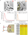
Behavior of colloidal Au and AgAu nanoparticles after incubation with S. aureus. Electron micrographs showing the accumulation of Au NPs at the bacterial cell surface but: a) no internalized NPs and NPs found inside only in the case of: b) AgAu alloy NPs. Nanoparticle size analysis of extra‐ and intracellularly located particles (Number‐weighted particle size distributions were derived from N individual particles located inside or outside the bacterial cells, x c values represent number‐mean values, and errors were calculated from the variance of the log‐normal fit functions) (c,d). Pathway of Au NPs and AgAu alloys schematically shown in (e). Elemental composition of intra‐ and extracellularly located particles after incubating S. aureus with AgAu NPs (f).
Figure 3
Figure 3
HAADF‐STEM micrograph of an S. aureus cell incubated with AgAu NPs. Visualization of particles close to the outer bacterial cell wall and particles located within the cell. Note that brighter contrast corresponds to heavier material (a). Elemental composition of selected extra‐(1) and intracellular (2) particles (b) extracted from EDS measurements. Proposed three‐staged REPER‐mechanism: (1) Extracellular dissolution and RElease of Ag+ cations from AgAu alloy NPs, (2) PEnetration of Ag+ cations into cells, and (3) intracellular Reduction to pure Ag NPs (c, step 3).

Similar articles

Cited by

References

    1. a) Cortie M. B., McDonagh A. M., Chem. Rev. 2011, 111, 3713; - PubMed
    2. b) Borah R., Verbruggen S. W., J. Phys. Chem. 2020, 124, 12081;
    3. c) Yang J., Shen D., Zhou L., Li W., Fan J., El‐Toni A. M., Zhang W.‐x., Zhang F., Zhao D., Adv. Healthcare Mater. 2014, 3, 1620. - PubMed
    1. a) Li L.‐L., Yin Q., Cheng J., Lu Y., Adv. Healthcare Mater. 2012, 1, 567; - PubMed
    2. b) Mazloomi‐Rezvani M., Salami‐Kalajahi M., Roghani‐Mamaqani H., Pirayesh A., Appl. Organomet. Chem. 2018, 32, 8;
    3. c) Indrasekara A. S. D. S., Paladini B. J., Naczynski D. J., Starovoytov V., Moghe P. V., Fabris L., Adv. Healthcare Mater. 2013, 2, 1370. - PubMed
    1. a) Chernousova S., Epple M., Angew. Chem. Int. Ed. Engl. 2013, 52, 1636; - PubMed
    2. b) Panicker S., Ahmady I. M., Han C., Chehimi M., Mohamed A. A., Mater. Today Chem. 2020, 16, 100237;
    3. c) Lv Z., He S., Wang Y., Zhu X., Adv. Healthcare Mater. 2021, 10, e2001806. - PubMed
    1. a) Taylor U., Tiedemann D., Rehbock C., Kues W. A., Barcikowski S., Rath D., Beilstein J. Nanotechnol. 2015, 6, 651; - PMC - PubMed
    2. b) Diem P. N. H., Phuong T. N. M., Hien N. Q., Quang D. T., Hoa T. T., Cuong N. D., J. Nanomater. 2020, 2020, 7195048.
    1. Ristig S., Chernousova S., Meyer‐Zaika W., Epple M., Beilstein J. Nanotechnol. 2015, 6, 1212. - PMC - PubMed

Publication types

LinkOut - more resources