. 2023 Nov;10(31):e2300473.
doi: 10.1002/advs.202300473. Epub 2023 Sep 3.

Photo-Chemical Stimulation of Neurons with Organic Semiconductors

Affiliations

Photo-Chemical Stimulation of Neurons with Organic Semiconductors

Achilleas Savva et al. Adv Sci (Weinh). 2023 Nov.

Abstract

Recent advances in light-responsive materials enabled the development of devices that can wirelessly activate tissue with light. Here it is shown that solution-processed organic heterojunctions can stimulate the activity of primary neurons at low intensities of light via photochemical reactions. The p-type semiconducting polymer PDCBT and the n-type semiconducting small molecule ITIC (a non-fullerene acceptor) are coated on glass supports, forming a p-n junction with high photosensitivity. Patch clamp measurements show that low-intensity white light is converted into a cue that triggers action potentials in primary cortical neurons. The study shows that neat organic semiconducting p-n bilayers can exchange photogenerated charges with oxygen and other chemical compounds in cell culture conditions. Through several controlled experimental conditions, photo-capacitive, photo-thermal, and direct hydrogen peroxide effects on neural function are excluded, with photochemical delivery being the possible mechanism. The profound advantages of low-intensity photo-chemical intervention with neuron electrophysiology pave the way for developing wireless light-based therapy based on emerging organic semiconductors.

Keywords: non-fullerene acceptors; organic bioelectronics; photo-stimulation.


Conflict of interest statement

The authors declare no conflict of interest.

Figures

Figure 1
Figure 1
Schematic representation of the photo‐stimulation of neurons in vitro using an organic pn heterojunction: PDCBT‐ITIC. Mouse cortical neurons were extracted and cultured directly on the photosensitive pn polymer junction generated by layer‐by‐layer type spin coating of each film. The absorption spectrum of the pn junction and the chemical structures of the semiconducting materials used are shown in the bottom panel.
Figure 2
Figure 2
a) A schematic of the photo‐EQCM‐D setup, used to record mass and photocurrent changes in‐operando. The thickness of the PDCBT‐ITIC films was calculated using the QCM‐D data (top graph) in dry conditions (solid line) and when they were immersed in PBS (dashed line). b) Mass changes of the PDCBT‐ITIC films and the corresponding photo‐current changes upon illumination. c) A schematic of the photodiode setup used to study the photo‐electrochemical operation of the PDCBT‐ITIC junctions in aqueous electrolytes. The photovoltage and photocurrent density response of the system is shown when the stimulation is conducted with a red light pulse (λ 630nm) with an intensity of 33 mW cm−2 for 10 ms and d) for 300 ms. e) An energy diagram showing the photodiode components and the photo‐electrochemical processes occurring upon light exposure, depicting photo‐capacitive charging at the semiconductor/electrolyte interface and photocathodic charge transfer to oxygen, producing hydrogen peroxide.
Figure 3
Figure 3
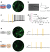
a) Live/Dead assay of mouse cortical neurons stained with calcein AM (green, live cells) and ethidium homodimer‐1 (red, dead cells) cultured on glass coverslips coated with polymer pn junctions and P‐d‐L. b) a picture of a neuron grown on a PDCBT‐ITIC coated glass coverslip during patch‐clamp measurements and the corresponding voltage time‐course trace, with injection of depolarization and hyperpolarization currents. c) Membrane potential recordings and action potential generation of neurons after several seconds of light exposure (yellow highlighted area). d) Raster plot, showing the action potential (black lines) generated by eleven individual neurons grown on polymer coated glass cover slips, right after light illumination. e) Live/Dead assays of the neurons grown on a glass/P‐d‐L substrate, and a representative membrane potential recording of such a neuron exposed to light illumination therein (n = 8). f) Live/Dead assays of the neurons grown on a glass cover slip coated with photoresist/P‐d‐L substrate, and a representative membrane potential recording of such a neuron exposed to light illumination (n = 3).
Figure 4
Figure 4
Membrane potential recordings of mouse cortical neurons exposed to different H2O2 concentrations in the dark. The cells were cultured on a) glass‐ P‐d‐L coverslips (n = 5) or b) glass ‐PDCBT‐ITIC‐P‐d‐L coverslips (n = 5).
Figure 5
Figure 5
a) Representative membrane potential traces of a mouse cortical neuron cultured on a glass coverslip half‐coated with PDCBT‐ITIC‐P‐d‐L. Primary neurons were seeded, maintained, and studied only on the bare glass‐P‐d‐L side. b) The traces were recorded from a culture cultured on top of glass ‐PDCBT‐ITIC‐P‐d‐L coverslip before and after the addition of HEPES to the culture. The highlighted areas represent the light pulses.

References

    1. Hamblin M. R., Nelson S. T., Strahan J. R., Photobiomodulation, Photomed., Laser Surg. 2018, 36, 241. - PMC - PubMed
    1. Chan W.‐M., Lam D. S. C., Lai T. Y. Y., Liu D. T. L., Li K. K. W., Yao Y., Wong T.‐H., Ophthalmology 2004, 111, 1576. - PubMed
    1. Maya‐Vetencourt J. F., Ghezzi D., Antognazza M. R., Colombo E., Mete M., Feyen P., Desii A., Buschiazzo A., Di Paolo M., Di Marco S., Ticconi F., Emionite L., Shmal D., Marini C., Donelli I., Freddi G., Maccarone R., Bisti S., Sambuceti G., Pertile G., Lanzani G., Benfenati F., Nat. Mater. 2017, 16, 681. - PMC - PubMed
    1. Berggren M., Głowacki E. D., Simon D. T., Stavrinidou E., Tybrandt K., Chem. Rev. 2022, 122, 4826. - PMC - PubMed
    1. Rohringer S., Holnthoner W., Chaudary S., Slezak P., Priglinger E., Strassl M., Pill K., Mühleder S., Redl H., Dungel P., Sci. Rep. 2017, 7, 10700. - PMC - PubMed

LinkOut - more resources