

Visualizing neutrophil extracellular traps in septic equine synovial and peritoneal fluid samples using immunofluorescence microscopy
- PMID: 37661696
- PMCID: PMC10621558
- DOI: 10.1177/10406387231196552
Visualizing neutrophil extracellular traps in septic equine synovial and peritoneal fluid samples using immunofluorescence microscopy
Abstract
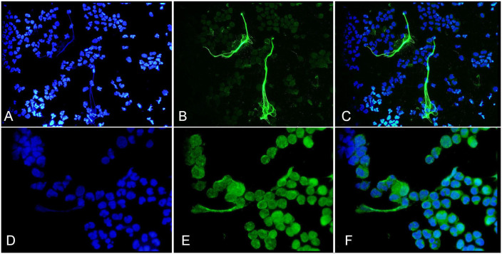
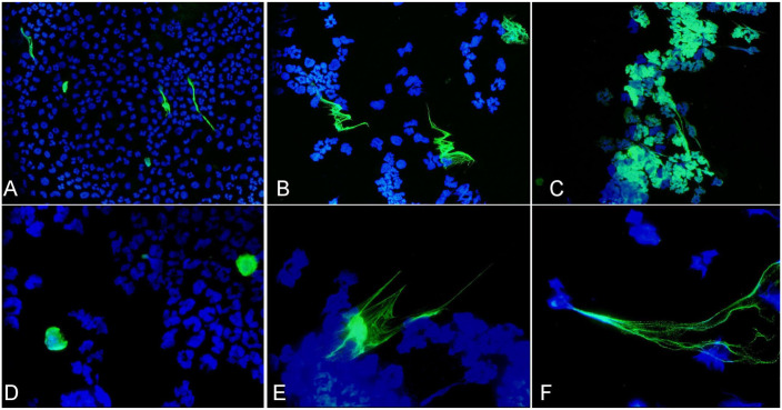
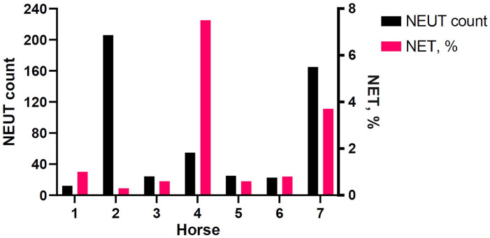
Septic synovitis and peritonitis are routinely diagnosed in horses based on clinical examination findings and laboratory assessment of synoviocentesis and abdominocentesis samples, respectively. Diagnosis is difficult in some cases because of an overlap in laboratory results for septic and non-septic inflammation. Neutrophil extracellular trap (NET) formation is part of the innate immune response against pathogens. Identifying and quantifying NETs, which have not been explored in clinical samples from horses with septic synovitis and peritonitis, to our knowledge, may be helpful in detecting infectious processes. Our main objective was to determine whether NETs could be visualized in septic equine synovial and peritoneal fluid cytology samples using immunofluorescence with antibodies against citrullinated histone H3 (Cit-H3) and myeloperoxidase (MPO). We analyzed 9 synovial and 4 peritoneal fluid samples. NET percentages were quantified using a simple counting technique, which is suitable for high-quality, well-preserved, and stained cytospin smears. NETs were evident in all septic samples and were absent in a non-septic sample; NETs were better visualized with Cit-H3 than with MPO immunolabeling. Overall, we believe that there is the potential for NETs and associated markers to be used to investigate and understand septic inflammation in horses.
Keywords: NETs; histones; horses; immunofluorescence; infection; peritoneal fluid; synovial fluid.
Conflict of interest statement
Declaration of conflicting interestsThe authors declared no conflicts of interest with respect to the research, authorship, or publication of this article.
Figures
Similar articles
-
A novel approach to identifying and quantifying neutrophil extracellular trap formation in septic dogs using immunofluorescence microscopy.BMC Vet Res. 2018 Jun 27;14(1):210. doi: 10.1186/s12917-018-1523-z. BMC Vet Res. 2018. PMID: 29945605 Free PMC article.
-
Detection of synovial sepsis in horses using enzymes as biomarkers.Equine Vet J. 2022 May;54(3):513-522. doi: 10.1111/evj.13459. Epub 2021 Jun 15. Equine Vet J. 2022. PMID: 33977535 Free PMC article.
-
Ex Vivo and In Vitro Analysis Identify a Detrimental Impact of Neutrophil Extracellular Traps on Eye Structures in Equine Recurrent Uveitis.Front Immunol. 2022 Feb 10;13:830871. doi: 10.3389/fimmu.2022.830871. eCollection 2022. Front Immunol. 2022. PMID: 35251020 Free PMC article.
-
Abdominocentesis techniques in horses.J Vet Emerg Crit Care (San Antonio). 2022 Jan;32(S1):72-80. doi: 10.1111/vec.13118. J Vet Emerg Crit Care (San Antonio). 2022. PMID: 35044064 Review.
-
Neutrophil Extracellular Traps in Asthma: Friends or Foes?Cells. 2022 Nov 7;11(21):3521. doi: 10.3390/cells11213521. Cells. 2022. PMID: 36359917 Free PMC article. Review.
References
-
- Alonso JM, et al.. Accuracy of differences in blood and peritoneal glucose to differentiate between septic and non-septic peritonitis in horses. Res Vet Sci 2020;132:237–242. - PubMed
MeSH terms
LinkOut - more resources
Full Text Sources
Research Materials
Miscellaneous
