. 2023 Nov 1;325(5):C1190-C1200.
doi: 10.1152/ajpcell.00166.2023. Epub 2023 Sep 4.

MARCH8 downregulation modulates profibrotic responses including myofibroblast differentiation

Affiliations

MARCH8 downregulation modulates profibrotic responses including myofibroblast differentiation

Xia Guo et al. Am J Physiol Cell Physiol. .

Abstract

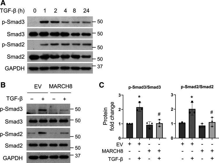
Interstitial lung diseases can result in poor patient outcomes, especially in idiopathic pulmonary fibrosis (IPF), a severe interstitial lung disease with unknown causes. The lack of treatment options requires further understanding of the pathological process/mediators. Membrane-associated RING-CH 8 (MARCH8) has been implicated in immune function regulation and inflammation, however, its role in the development of pulmonary fibrosis and particularly the fibroblast to myofibroblast transition (FMT) remains a gap in existing knowledge. In this study, we demonstrated decreased MARCH8 expression in patients with IPF compared with non-PF controls and in bleomycin-induced PF. TGF-β dose- and time-dependently decreased MARCH8 expression in normal and IPF human lung fibroblast (HLFs), along with induction of FMT markers α-SMA, collagen type I (Col-1), and fibronectin (FN). Interestingly, overexpression of MARCH8 significantly suppressed TGF-β-induced expression of α-SMA, Col-1, and FN. By contrast, the knockdown of MARCH8 using siRNA upregulated basal expression of α-SMA/Col-1/FN. Moreover, MARCH8 knockdown enhanced TGF-β-induced FMT marker expression. These data clearly show that MARCH8 is a critical "brake" for FMT and potentially affects PF. We further found that TGF-β suppressed MARCH8 mRNA expression and the proteasome inhibitor MG132 failed to block MARCH8 decrease induced by TGF-β. Conversely, TGF-β decreases mRNA levels of MARCH8 in a dose- and time-dependent manner, suggesting the transcriptional regulation of MARCH8 by TGF-β. Mechanistically, MARCH8 overexpression suppressed TGF-β-induced Smad2/3 phosphorylation, which may account for the observed effects. Taken together, this study demonstrated an unrecognized role of MARCH8 in negatively regulating FMT and profibrogenic responses relevant to interstitial lung diseases.NEW & NOTEWORTHY MARCH8 is an important modulator of inflammation, immunity, and other cellular processes. We found that MARCH8 expression is downregulated in the lungs of patients with idiopathic pulmonary fibrosis (IPF) and experimental models of pulmonary fibrosis. Furthermore, TGF-β1 decreases MARCH8 transcriptionally in human lung fibroblasts (HLFs). MARCH8 overexpression blunts TGF-β1-induced fibroblast to myofibroblast transition while knockdown of MARCH8 drives this profibrotic change in HLFs. The findings support further exploration of MARCH8 as a novel target in IPF.

Keywords: IPF; MARCH8; TGF-β; fibroblast; lung.


Conflict of interest statement

No conflicts of interest, financial or otherwise, are declared by the authors.

Figures

None
Graphical abstract
Figure 1.
Figure 1.
MARCH8 expression is downregulated in patients with idiopathic pulmonary fibrosis (IPF). A: representative Masson’s trichrome staining of lung tissues from histologically normal donors and patients with IPF showing increased collagen deposition (blue) in patients with IPF compared with that of controls. B: immunohistochemical (IHC) staining of MARCH8 showed that compared with the notable expression of MARCH8 in the control lung tissue, MARCH8 expression was much decreased. Scale bars represent 100 μm, images were taken at the same magnification. Boxes show enlarged views of the areas of interest. C: reciprocal intensity method was used for the quantification of MARCH8 staining as in B from 10 views per slide/donor, n = 3 donors/group; a.u., arbitrary units. *P < 0.05 compared with normal control, tested by unpaired Student’s t test.
Figure 2.
Figure 2.
MARCH8 expression is decreased in preclinical models of pulmonary fibrosis. A: representative immunohistochemical (IHC) staining of MARCH8 in the lungs of C57BL/6 mice received intratracheal instillation of saline or bleomycin for 21 days. Black boxes indicate the enlarged view of areas of interest. Bar represents 100 µm. B: reciprocal intensity method was used for the quantification of MARCH8 as in a form of 10 views per slide/mouse, n = 5 mice/group. *P < 0.05 compared with saline group, tested by unpaired Student’s t test; a.u., arbitrary units. C: representative IHC images of the lung tissues from C57BL/6 mice administered adenoviral control (control) or TGF-β1 adenovirus (TGF-β) intratracheally for 14 days. Black boxes indicate the enlarged view of areas of interest. Bar represents 100 µm. D: reciprocal intensity method was used for the quantification of MARCH8 as in C from 10 views per slide/mouse, n = 5 mice/group. *P < 0.05 compared with adenoviral control group, tested by unpaired Student’s t test; a.u., arbitrary units. E: lung tissues were collected from saline and Bleomycin treated mice for Western blot analysis of MARCH8. Actin was used as the loading control. n = 4 mice/group. F: ImageJ was used to quantify the band density of MARCH8 as shown in E and adjusted to the corresponding loading control. The relative values were used to generate the graph. *P < 0.05 compared with saline group, tested by unpaired Student’s t test. n = 4 mice/group.
Figure 3.
Figure 3.
MARCH8 expression was decreased by transforming growth factor-beta (TGF-β) in primary normal (A, B, E) and idiopathic pulmonary fibrosis (IPF; C, D, F) human lung fibroblasts (HLFs). A: serum-starved primary normal HLFs were treated with different doses of TGF-β for 24 h, followed by Western blotting (WB) analysis of MARCH8 and fibroblast to myofibroblast transition (FMT) markers including alpha-smooth muscle actin (α-SMA), fibronectin (FN), and collagen type I (Col-1). GAPDH was used as the loading control. B: quantification of A based on 3 independent experiments. C: serum-starved primary IPF HLFs were treated with different doses of TGF-β, followed by WB analysis of MARCH8 and FMT markers. D: the quantification of C based on 3 independent experiments. E and F: serum-starved primary normal (E) and IPF (F) HLFs were treated with 5 ng/mL TGF-β for different times as indicated, followed by WB analysis of given proteins. NIH ImageJ software was used to quantify the band intensity. *P < 0.05 compared with vehicle control, n = 3 independent experiments. One-way ANOVA followed by Dunnett’s test was used for multiple comparisons.
Figure 4.
Figure 4.
Overexpression of MARCH8 attenuates transforming growth factor-beta (TGF-β)-induced fibroblast to myofibroblast transition (FMT) in primary normal human lung fibroblasts (HLFs). A: primary HLFs were transfected with empty vector control (EV) or MARCH8 plasmid for 48 h and MARCH8 was detected by Western blotting (WB) with GAPDH as the loading control. B: primary HLFs were transfected with EV or MARCH8 plasmid, followed by treatment with TGF-β (5 ng/mL) for an additional 24 h. Whole cell lysates were collected for WB analysis of MARCH8 and FMT markers (α-SMA, Col-1, and FN). GAPDH was used as the loading control. C: quantification of MARCH8 and FMT markers as in B, based on three independent experiments. NIH ImageJ software was used to quantify the band intensity. *P < 0.05 vs. control EV group; #P < 0.05 vs. TGF-β-treated EV control, n = 3 independent experiments. One-way ANOVA followed by Dunnett’s test was used for multiple comparisons. α-SMA, alpha-smooth muscle actin; Col-1, collagen 1A1; FN, fibronectin.
Figure 5.
Figure 5.
Inhibition of MARCH8 promotes fibroblast to myofibroblast transition (FMT) in primary normal human lung fibroblasts (HLFs). A: primary normal HLFs were transfected with scramble or siRNA targeting MARCH8 (siMARCH8) and whole cell lysates were collected 48 h after transfection for Western blotting (WB) analysis of MARCH8 and FMT markers (α-SMA, Col-1, and FN). β-actin was used as the loading control. B: quantification of MARCH8 and FMT markers as in A, based on three independent experiments. *P < 0.05 vs. scramble group based on three independent experiments tested by unpaired Student’s t test. C: primary normal HLFs were transfected with scramble or siMARCH8, followed by treatment with TGF-β (5 ng/mL) for an additional 24 h. Whole cell lysates were collected for WB analysis of MARCH8 and FMT markers. D: quantification of MARCH8 and FMT markers as in C, based on three independent experiments. NIH ImageJ software was used to quantify the band intensity. *P < 0.05 vs. scramble control; #P < 0.05 vs. TGF-β-treated scramble group, n = 3 independent experiments. One-way ANOVA followed by Dunnett’s test was used for multiple comparisons. α-SMA, alpha-smooth muscle actin; Col-1, collagen 1A1; FN, fibronectin.
Figure 6.
Figure 6.
Overexpression of MARCH8 inhibits the activation of Smad3 and Smad2 induced by transforming growth factor-beta (TGF-β). A: serum-starved primary normal human lung fibroblasts (HLFs) were treated with TGF-β (5 ng/mL), and whole cell lysates were collected at various time points to detect phosphor-Smad3 (p-Smad3) and p-Smad2 by Western blotting (WB). GAPDH was used as the loading control. B: primary normal HLFs were transfected with control empty vector (EV) or MARCH8 plasmid (MARCH8), followed by treatment with TGF-β (5 ng/mL) for an additional 2 h. Whole cell lysates were collected for WB analysis of p-Smad3, Smad3, p-Smad2, and Smad2. GAPDH was used as the loading control. C: quantification of the ratios of p-Smad3/Smad3 and p-Smad2/Smad2 as in B, based on three independent experiments. NIH ImageJ software was used to quantify the band intensity. *P < 0.05 vs. EV control group; #P < 0.05 vs. TGF-β-treated EV group, n = 3 independent experiments. One-way ANOVA followed by Dunnett’s test was used for multiple comparisons.
Figure 7.
Figure 7.
Transforming growth factor-beta (TGF-β) downregulates MARCH8 at the transcriptional level. A-B, qPCR analysis showed that MARCH8 mRNA was reduced by TGF-β in dose-dependent (A, 12 h) and time-dependent (B, 5 ng/mL) manners in primary normal human lung fibroblasts (HLFs). GAPDH was used as the internal control, n = 3 replicates. *P < 0.05 vs. vehicle control group. C: primary normal HLFs were pretreated with actinomycin D (Act D, 1 μg/mL) for 30 min, followed by TGF-β treatment (5 ng/mL, 16 h) for Western blotting (WB) analysis of MARCH8. GAPDH was the loading control. D: the quantification of C based on three independent experiments. *P < 0.05. NS, not significant. E: primary normal HLFs were pretreated with the proteasome inhibitor MG132 (5 μM) for 30 min, followed by TGF-β treatment (5 ng/mL, 16 h) for WB analysis of MARCH8. F: the quantification of E based on three independent experiments. *P < 0.05 based on three independent experiments. G: primary normal HLFs were treated with TGF-β (5 ng/mL, 16 h), followed by 10 μg/mL cycloheximide (CHX) treatment for the indicated times. Whole cell lysates were collected for WB analysis of MARCH8 degradation. H: MARCH8 levels were plotted relative to those at time 0 of CHX treatment (G) after being quantified by NIH ImageJ software and normalized to Actin. Solid line indicates control group and dash line indicates TGF-β group. One-way ANOVA followed by Dunnett’s test was used for multiple comparisons.
Figure 8.
Figure 8.
MARCH8 downregulation by transforming growth factor-beta (TGF-β) is p38 MAPK pathway dependent. A: primary normal HLFs were pretreated with various inhibitors (10 μM) of Smad3 (SIS3), AKT (Ly294002), ERK (U0126), JNK (SP600125), and p38 MAPK (SB203580) for 30 min, followed by TGF-β treatment (5 ng/mL) for an additional 16 h. Whole cell lysates were collected for Western blotting (WB) analysis of MARCH8 with GAPDH as a loading control. B: the quantification of MARCH8 as in A, based on three independent experiments. *P < 0.05 vs. TGF-β alone group, n = 3 independent experiments. One-way ANOVA followed by Dunnett’s test was used for multiple comparisons. C: primary normal human lung fibroblasts (HLFs) were pretreated with various concentrations of SB203580 for 30 min, followed by treatment with 5 ng/mL TGF-β for an additional 16 h. Whole cell lysates were collected for WB analysis of MARCH8 with GAPDH as a loading control. D: the quantification of MARCH8 as shown in C, based on three independent experiments. NIH ImageJ software was used to quantify the band intensity. *P < 0.05 vs. TGF-β alone group, n = 3 independent experiments. One-way ANOVA followed by Dunnett’s test was used for multiple comparisons.

References

    1. Martinez FJ, Collard HR, Pardo A, Raghu G, Richeldi L, Selman M, Swigris JJ, Taniguchi H, Wells AU. Idiopathic pulmonary fibrosis. Nat Rev Dis Primers 3: 17074, 2017. doi:10.1038/nrdp.2017.74. - DOI - PubMed
    1. Park Y, Ahn C, Kim TH. Occupational and environmental risk factors of idiopathic pulmonary fibrosis: a systematic review and meta-analyses. Sci Rep 11: 4318, 2021. doi:10.1038/s41598-021-81591-z. - DOI - PMC - PubMed
    1. Raghu G, Collard HR, Egan JJ, Martinez FJ, Behr J, Brown KK, Colby TV, Cordier JF, Flaherty KR, Lasky JA, Lynch DA, Ryu JH, Swigris JJ, Wells AU, Ancochea J, Bouros D, Carvalho C, Costabel U, Ebina M, Hansell DM, Johkoh T, Kim DS, King TE Jr, Kondoh Y, Myers J, Müller NL, Nicholson AG, Richeldi L, Selman M, Dudden RF, Griss BS, Protzko SL, Schünemann HJ; ATS/ERS/JRS/ALAT Committee on Idiopathic Pulmonary Fibrosis. An official ATS/ERS/JRS/ALAT statement: idiopathic pulmonary fibrosis: evidence-based guidelines for diagnosis and management. Am J Respir Crit Care Med 183: 788–824, 2011. doi:10.1164/rccm.2009-040GL. - DOI - PMC - PubMed
    1. Sauleda J, Núñez B, Sala E, Soriano JB. Idiopathic pulmonary fibrosis: epidemiology, natural history, phenotypes. Med Sci (Basel) 6: 110, 2018. doi:10.3390/medsci6040110. - DOI - PMC - PubMed
    1. Wolters PJ, Collard HR, Jones KD. Pathogenesis of idiopathic pulmonary fibrosis. Annu Rev Pathol 9: 157–179, 2014. doi:10.1146/annurev-pathol-012513-104706. - DOI - PMC - PubMed

Publication types

MeSH terms

LinkOut - more resources