Case Reports
. 2023 Aug 29:16:491-496.
doi: 10.2147/IMCRJ.S416492. eCollection 2023.

COVID-19 Related Acute Macular Neuroretinopathy (AMN): A Case Series

Affiliations
Case Reports

COVID-19 Related Acute Macular Neuroretinopathy (AMN): A Case Series

Truong Andy Vu et al. Int Med Case Rep J. .

Abstract

Purpose: Following the emergence of coronavirus disease 2019 (COVID-19) eye care practitioners have become accustomed to identifying and managing an array of ocular complications following the viral infection. Acute macular neuroretinopathy (AMN) is one such complication that has been reported. While the etiology of AMN has eluded researchers, current literature is suggestive of a microvascular compromise within the deep capillary plexus of the retina.

Observations: In this case series, we aim to explore two individual cases of presumed AMN following confirmed COVID-19 infection. Our observations and findings support the diagnosis of AMN following the criteria outlined in literature.

Conclusion and importance: Although acute macular neuroretinopathy is rare, it should be considered by clinicians when considering diagnosis. With the changing landscape of the pandemic, careful and thorough history and testing are key in the diagnosis of AMN.

Keywords: COVID-19; acute macular retinopathy; neuroretinopathy.


Conflict of interest statement

The following authors have no financial disclosures: T.V, M.S, S.M, W.S

Figures

Figure 1
Figure 1
(A) 10–2 Visual field (VF): OD mild scattered VF depression. (B) 10–2 Visual field (VF): OS mild scattered VF depression and scotoma. Arrow pointing to a wedge-shaped scotoma.
Figure 2
Figure 2
(A) Optical coherence tomography (OCT): OD slight disruption of nasal parafoveal outer photoreceptor segments and interdigitation zone. (B) Optical coherence tomography (OCT): OS focal atrophy and excavation of nasal parafoveal outer photoreceptor segments and interdigitation zone. Blue arrow: Outer nuclear layer; Red arrow: Photoreceptors; Green arrow: Outer plexiform layer.
Figure 3
Figure 3
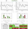
Visual evoked potential: Decreased amplitude with high contrast test stimuli OD and increased latency of high contrast test stimuli OS. Red color cells outside normative ranges. Green cells are within normative ranges.
Figure 4
Figure 4
(A) 10–2 Visual field (VF): OD mild scattered VF depression. (B) 10–2 Visual field (VF): OS mild scattered VF depression and temporal scotoma in the left eye. Arrow pointing to a wedge-shaped scotoma.
Figure 5
Figure 5
(A) Optical coherence tomography (OCT) ganglion cell analysis: Normal macular profile OD. Significance mapping: Green is p>5%, yellow is p<5% and red is <1%. Thickness mapping: Blue 50 to ~ 80 microns, green >80 to ~180 microns, yellow >180 to <175 microns. (B) Optical coherence tomography (OCT) ganglion cell analysis: Atrophy of nasal papillary bundle OS. Significance mapping: Green is p>5%, yellow is p<5% and red is <1%. Thickness mapping: Blue 50 to ~ 80 microns, green >80 to ~180 microns, yellow >180 to <175 microns. (C) Optical coherence tomography (OCT) Full thickness analysis: Atrophy of nasal papillary bundle OS. Thickness mapping: Blue 50 to ~ 160 microns, green 200 to ~300 microns, yellow >300 to <370 microns. Deviation mapping: Red: ~25 to 40%, Orange: ~20 to 25%, yellow: −10 to 15%, green −5 to 5%, blue −25 to −15%, black and grey −30 to −50%. (D) Optical coherence tomography (OCT): OD 5 months follow up shows normal macular scan. (E) Optical coherence tomography (OCT): OS macular scan 5 months shows minimal improved architecture of ellipsoid zone, photoreceptor outer segments and interdigitation zone. Blue arrow: Outer nuclear layer; Red arrow: Photoreceptors; Green arrow: Outer plexiform layer.

References

    1. Bos PJM, Deutman AF. Acute Macular Neuroretinopathy. Am J Ophthalmol. 1975;80(4):573–584. doi:10.1016/0002-9394(75)90387-6 - DOI - PubMed
    1. El-Asrar AM A, Herbort CP, Tabbara KF. Differential Diagnosis of Retinal Vasculitis. Middle East Afr J Ophthalmol. 2009;16(4):202–218. doi:10.4103/0974-9233.58423 - DOI - PMC - PubMed
    1. Rahimy E, Sarraf D. Paracentral acute middle maculopathy spectral-domain optical coherence tomography feature of deep capillary ischemia. Curr Opin Ophthalmol. 2014;25(3):207. doi:10.1097/ICU.0000000000000045 - DOI - PubMed
    1. Bhavsar KV, Lin S, Rahimy E, et al. Acute macular neuroretinopathy: a comprehensive review of the literature. Surv Ophthalmol. 2016;61(5):538–565. doi:10.1016/j.survophthal.2016.03.003 - DOI - PubMed
    1. Jalink MB, Bronkhorst IHG. A sudden rise of patients with acute macular neuroretinopathy during the COVID-19 pandemic. Case Rep Ophthalmol. 2022;13:96–103. doi:10.1159/000522080 - DOI - PMC - PubMed

Publication types