[Preprint]. 2023 Aug 22:2023.08.21.23294276.
doi: 10.1101/2023.08.21.23294276.

Insights into the Mechanisms and Structure of Breakage-Fusion-Bridge Cycles in Cervical Cancer using Long-Read Sequencing

Affiliations

Insights into the Mechanisms and Structure of Breakage-Fusion-Bridge Cycles in Cervical Cancer using Long-Read Sequencing

Isabel Rodriguez et al. medRxiv. .

Update in

Abstract

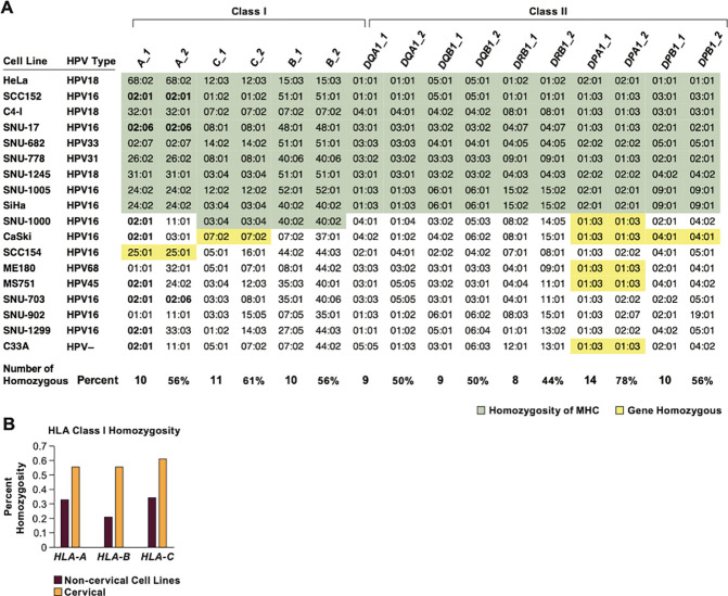
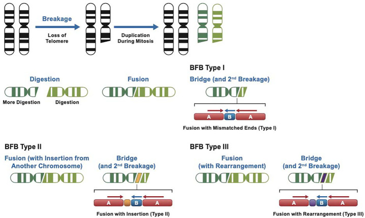
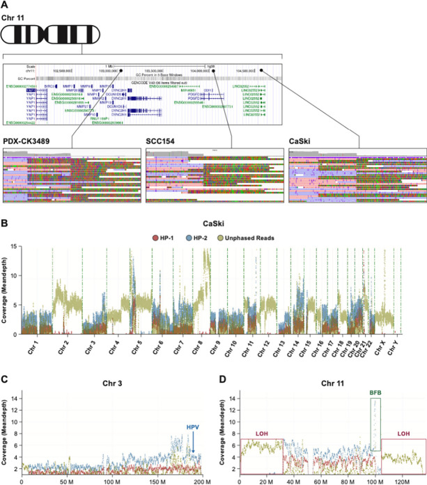
Cervical cancer is caused by human papillomavirus (HPV) infection, has few approved targeted therapeutics, and is the most common cause of cancer death in low-resource countries. We characterized 19 cervical and four head and neck cell lines using long-read DNA and RNA sequencing and identified the HPV types, HPV integration sites, chromosomal alterations, and cancer driver mutations. Structural variation analysis revealed telomeric deletions associated with DNA inversions resulting from breakage-fusion-bridge (BFB) cycles. BFB is a common mechanism of chromosomal alterations in cancer, and this is one of the first analyses of these events using long-read sequencing. Analysis of the inversion sites revealed staggered ends consistent with exonuclease digestion of the DNA after breakage. Some BFB events are complex, involving inter- or intra-chromosomal insertions or rearrangements. None of the BFB breakpoints had telomere sequences added to resolve the dicentric chromosomes and only one BFB breakpoint showed chromothripsis. Five cell lines have a Chr11q BFB event, with YAP1/BIRC2/BIRC3 gene amplification. Indeed, YAP1 amplification is associated with a 10-year earlier age of diagnosis of cervical cancer and is three times more common in African American women. This suggests that cervical cancer patients with YAP1/BIRC2/BIRC3-amplification, especially those of African American ancestry, might benefit from targeted therapy. In summary, we uncovered new insights into the mechanisms and consequences of BFB cycles in cervical cancer using long-read sequencing.

Keywords: HPV integration; Human Papillomavirus; breakage-bridge-fusion events; cervical cancer; extrachromosomal DNA.


Figures

Figure 1.
Figure 1.. Cell line patient demographics and HPV expression data.
A. The distribution of the ancestry of the subjects, HPV types, and cancer and histological types among the 22 cell lines is shown. B. The cancer driver gene mutations found mutated in at least one cell line are shown. Orange highlights are pathogenic variants and in yellow are variants of unknown impact; H, homozygous variants; WT, wild type; AMP, amplification; no CNV, no copy number variants were found. C. The cDNA or RNA reads per million reads mapped (RPM) to the HPV genome is shown. D. The percentage of HPV E6/E7 containing reads that are unspliced in the E6 gene.
Figure 2.
Figure 2.. HLA class I homozygosity in cervical cancer cell lines
A. The genotype for each class I and II HLA genes is shown with loci homozygous across all or most genes indicated in green, and across 1–3 genes in yellow. The number and percent homozygosity for each gene is indicated at the bottom. B. The percent of tumor samples homozygous for individual HLA class I genes. The data for non-cervical cancer cell lines are from (Boegel et al., 2014).
Figure 3:
Figure 3:. HPV integration and gene expression.
A. A map of all integration events in the human genome is shown for 20 HPV+ cell lines. Each point represents an integration event, of which there may be one or multiple integration breakpoints. For the cell lines CaSki and SCC152, with multiple integrations, only the transcriptionally active site is shown. Each point lies in an approximate location of the integration event. The exact locations are included in Table 1 and Supplementary Table 2. B. Diagram of the HPV integration locus and flanking genes in four cell lines. The red arrows indicate a gene at the integration locus that is overexpressed in that cell line. C. The relative gene expression of each gene at or near an integration site was calculated as a Z-score; bold boxes are genes that are near the HPV integration. Darker green colors indicate higher values, showing those genes are highly expressed in those cell lines.
Figure 4.
Figure 4.. TP53 and RB1 mutations in HPV-negative and moderate-risk HPV cell lines.
A. The Integration site of HPV30 in HT-3 cells demonstrates that the integration is 19 kb 3’ to the RB1 gene within the RCBTB2 gene. B. A model showing that the HPV E6 and E7 proteins inhibit p53 and pRB respectively and mutations in RB1 and TP53 occur almost exclusively in cell lines without HPV or HPV of unknown risk.
Figure 5.
Figure 5.. Structure of BFB events.
A. The alignment of reads at the inversion site of a Type I BFB event on Chr17q in HT-3 cells is shown. The coverage drops from 95x (segment A) to 54x (segment B) to 16-fold (reads from non-rearranged allele). All soft-clipped portions of reads are inverted in relation to the aligned portion of the reads. Arrows mark the start and end of the segment shown as B in the diagram below. All the reads spanning the junction are consistent with the fusion of inverted chromatids with staggered ends. B. A proposed model for the formation of the BFB junction of chromosome 2 in SNU-1000 cells, a Type II BFB event. Following the deletion of one of the ends (del 1052 bp), a segment of chromosome 7 is inserted in between the joined chromosomes. The coverage drops from 52x (segment A) to 33x (segment B) to 11x. C. A Type III BFB event on chromosome 11p in SNU-682 cells, inside the WT1 gene. The coverage drops from 75x to 6x. The junction contains a complex sequence likely derived from sequences adjacent to the breakpoint (see Supplemental Figure 4G).
Figure 6.
Figure 6.. Model for BFB types.
Model for a BFB Type I event. A telomere deletion results in a pair of deleted chromatids during mitosis. The free ends are subject to exonuclease digestion, and uneven digestion generates staggered ends. Fusion results in a lower copy number (one half) of the B segment. Type II events are formed when a segment from another chromosome (orange) is inserted at the junction, and Type III events involve insertion of a sequence derived from the sequences flanking the breakage site (purple).
Figure 7.
Figure 7.. Map of the region of chromosome 11q22 with a cluster of 3 BFB events.
A. Map of the chromosome 11q22 region containing the YAP1, BIRC2, and BIRC3 genes is shown along with the location of three BFB events, in independent models. Detail of the read coverage aligned to HG38 is shown for the HNSCC PDX line (PDX-CK3489), SCC154 HNSCC cells and CaSki cervical cancer cells. All soft-clipped reads are inverted in relation to the aligned forward (blue) and reverse (pink) read segments. B. Phase reads copy number plot of the CaSki genome displaying reads assigned to haplotype 1 (HP-1) and 2 (HP-2) as well as unphased reads. Large blocks of unphased reads are due to loss-of-heterozygosity (LOH). C. Detail of chromosome 3 is shown indicating the location of multiple HPV integrations on 3q22–27 (131–188 MB). D. Plot of chromosome 11 showing the phased amplification of the YAP1 region (BFB) and the deletion of one haplotype and LOH after the BFB event extending to the telomere.
Figure 8.
Figure 8.. Analysis of YAP1 gene amplification in 31 cervical tumors.
A. Copy number data for genes centromeric and telomeric to YAP1 in all TCGA cervical tumors with Log2 copy number values for YAP1 >1. The location of the YAP1, BIRC2 and BIRC3 genes are highlighted. The location of each gene in MB is given below the gene symbol. B. Comparison of the age of diagnosis between women with YAP1-amplified and unamplified tumors C. Self-identified race of cervical cancer patients with and without YAP1 amplification. Data from TCGA, MSKCC metastatic cancer, and AACR GENIE cohort were obtained from cbioportal (https://www.cbioportal.org) (Cerami et al., 2012; Gao et al., 2013).

References

    1. Akagi K., Li J., Broutian T. R., Padilla-Nash H., Xiao W., Jiang B., Rocco J. W., Teknos T. N., Kumar B., Wangsa D., et al. (2014). Genome-wide analysis of HPV integration in human cancers reveals recurrent, focal genomic instability. Genome Res 24, 185–199. - PMC - PubMed
    1. Andre F., Ciruelos E., Rubovszky G., Campone M., Loibl S., Rugo H. S., Iwata H., Conte P., Mayer I. A., Kaufman B., et al. (2019). Alpelisib for PIK3CA-Mutated, Hormone Receptor-Positive Advanced Breast Cancer. N Engl J Med 380, 1929–1940. - PubMed
    1. Bagutti C., Forro G., Ferralli J., Rubin B., and Chiquet-Ehrismann R. (2003). The intracellular domain of teneurin-2 has a nuclear function and represses zic-1-mediated transcription. J Cell Sci 116, 2957–2966. - PubMed
    1. Bailey M. H., Tokheim C., Porta-Pardo E., Sengupta S., Bertrand D., Weerasinghe A., Colaprico A., Wendl M. C., Kim J., Reardon B., et al. (2018). Comprehensive Characterization of Cancer Driver Genes and Mutations. Cell 173, 371–385 e318. - PMC - PubMed
    1. Berger A. C., Korkut A., Kanchi R. S., Hegde A. M., Lenoir W., Liu W., Liu Y., Fan H., Shen H., Ravikumar V., et al. (2018). A Comprehensive Pan-Cancer Molecular Study of Gynecologic and Breast Cancers. Cancer Cell 33, 690–705 e699. - PMC - PubMed

Publication types

LinkOut - more resources