[Preprint]. 2023 Aug 24:2023.08.23.554128.
doi: 10.1101/2023.08.23.554128.

Antibodies Expand the Scope of Angiotensin Receptor Pharmacology

Affiliations

Antibodies Expand the Scope of Angiotensin Receptor Pharmacology

Meredith A Skiba et al. bioRxiv. .

Update in

  • Antibodies expand the scope of angiotensin receptor pharmacology.
    Skiba MA, Sterling SM, Rawson S, Zhang S, Xu H, Jiang H, Nemeth GR, Gilman MSA, Hurley JD, Shen P, Staus DP, Kim J, McMahon C, Lehtinen MK, Rockman HA, Barth P, Wingler LM, Kruse AC. Skiba MA, et al. Nat Chem Biol. 2024 Dec;20(12):1577-1585. doi: 10.1038/s41589-024-01620-6. Epub 2024 May 14. Nat Chem Biol. 2024. PMID: 38744986 Free PMC article.

Abstract

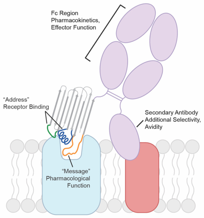
G protein-coupled receptors (GPCRs) are key regulators of human physiology and are the targets of many small molecule research compounds and therapeutic drugs. While most of these ligands bind to their target GPCR with high affinity, selectivity is often limited at the receptor, tissue, and cellular level. Antibodies have the potential to address these limitations but their properties as GPCR ligands remain poorly characterized. Here, using protein engineering, pharmacological assays, and structural studies, we develop maternally selective heavy chain-only antibody ("nanobody") antagonists against the angiotensin II type I receptor (AT1R) and uncover the unusual molecular basis of their receptor antagonism. We further show that our nanobodies can simultaneously bind to AT1R with specific small-molecule antagonists and demonstrate that ligand selectivity can be readily tuned. Our work illustrates that antibody fragments can exhibit rich and evolvable pharmacology, attesting to their potential as next-generation GPCR modulators.


Conflict of interest statement

Competing Interests A.C.K., C.M., L.W., D.P.S., and M.A.S., are co-inventors on patent application for AT1R blocking nanobodies. A.C.K. is a cofounder and consultant for biotechnology companies Tectonic Therapeutic and Seismic Therapeutic, and also for the Institute for Protein Innovation, a nonprofit research institute. L.M.W. is a scientific advisor for Septerna. D.P.S. is a Septerna employee. C.M. is a Sanofi employee.

Figures

Figure 1.
Figure 1.
Evolution of AT118 family of nanobody antagonists. a) Structure of AT118-H (PDB 7T83) and CDR sequences of nanobodies described in this study colored by CDR. Shaded positions vary between nanobodies. Full nanobody sequences are provided in Supplementary Table 1. b) Sites substituted in AT118-H library. Variant enrichment was tracked through each selection round via deep sequencing. c) AT118-H and evolved variant AT118-L displace olmesartan from cell membranes containing AT1R. Error bars represent mean ± standard error from three experiments. d) AT118-L Fc fusion proteins lacking the ability to interact with FcRn (pMAS493, Supplementary Table 1) do not accumulate in fetal serum of mice compared to AT118-L Fc fusion proteins with a WT Fc (pMAS430, Supplementary Table 1). Error bars represent mean ± standard error from twelve embryos from three separate litters.
Fig 2.
Fig 2.. Structure of H bound to AT1R.
a) Cryo-EM map colored by component: AT118-H (gray), AT1R (light blue), BRIL (purple), anti-BRIL Fab (light pink), anti-Fab nanobody (dark pink) b) Model of the AT118-H AT1R complex from local refinement maps. Image is colored as in a) with extracellular loops and AT118-H CDRs colored as follows: ECL1 (yellow), ECL2 (light pink), ECL3 (red), CDR1 (dark blue), CDR2 (green), CDR3 (orange).
Fig 3.
Fig 3.. AT118-H mimics peptide agonist binding to AT1R.
a) Surface rendering of AT118-H bound to AT1R as colored in Fig 2b. CDR3 (orange) engages with the orthosteric pocket of AT1R. b) surface rendering of active-state AT1R bound to the endogenous peptide agonist AngII (gray) (PDB 6OS0). AngII engages deeply in the orthosteric pocket. c) AT118-H binding is driven by engagement of CDRs 2 (green) and 3 (orange) with ECL2 (pink). Dashed lines indicate hydrogen bonds. d) AT118-H CDR3 (orange) forms β-strand and hydrophobic interactions with ECL2 (pink). The hydrophobic network is further supported by residues on ECL1 and TM2. e) AngII (gray sticks) and CDR3 share similar hydrophobic interactions with ECL2 (pink). f) In peptide agonist bound structures, the N-terminus of AT1R stabilizes the position of ECL2 through β-strand interactions. g) Binding of AT118-H to Expi293 cells expressing AT1R variants. Error bars represent mean ± standard error from three experiments. In the AT2R N-terminus chimera residues 2-16 of AT1R are replaced with residues of 3-33 of human AT2R. The AT2R ECL1 chimera contains M90Y, E91R, R93D, and P95L substitution and ECL3 chimera contains I270V, R272N, D273S substitutions.
Fig 4.
Fig 4.. AT118-H stabilizes a hybrid active-inactive state of AT1R.
a-b) R31CDR1 (a, blue sticks) and R99CDR3 (a, orange sticks) of AT118-H mimic R2 of AngII (b, gray sticks) positioning the extracellular side of Asp rich TMs 6 and 7. c) TM6, ECL3, and TM7 are displaced outward in AT118-H (light blue) and AngII (green) bound AT1R compared to small-molecule antagonist bound AT1R (purple) (PDB 4ZUD). d) AT118-H partially engages the allosteric activation network between AT1R’s orthosteric pocket and the intracellular side of AT1R. Switches indicated with the following arrows are activated (black), partially activated (dashed lines), unengaged (gray). e) AT118-H induced movements of TM5 and TM6 (light blue) induces inward movement of K1195.42 and downward rotation of W2536.48 compared to the inactive state (purple). f) W2536.48 does not fully rotate into its active-state position (green) and cannot displace N1113.35 and L1123.36 g) Partial movement of W2536.48 repositions Y2927.43 and F2085.51 from the canonical inactive state (purple) to h) active state positions (green), but is not sufficient to rotate the F2496.44 and F2506.45 rachet and displace TM6 to open up the intracellular effector binding site.
Fig 5.
Fig 5.. AT118 family members exhibit probe dependence with small molecules.
a) AT118-H and the evolved AT118-L variant accommodate binding of the small-molecule antagonist losartan (gray sticks). AT118-L CDR3 (purple) engages deeper within the orthosteric pocket compared to AT118-H (orange). b) S102CDR3 of AT118-H and c) D101CDR3 of AT118-L controls the depth of CDR3 within the orthosteric pocket through interactions with TM7. d) The backbone carbonyl of S102CDR3 of AT118-L is within hydrogen binding distance of the alcohol substituent (R1 in e) of losartan (gray sticks). e) Olmesartan and losartan derivatives vary in substituents on the imidazole ring. f) Probe dependent binding of AT118-H (orange) and AT118-L (purple) with losartan and olmesartan derivatives. Error bars represent mean ± standard error from three experiments. g) CDR3 of AT118-L clashes with the carboxylic acid (R1 in e) of modeled olmesartan (green sticks, modeled based upon PDB: 4ZUD).
Fig 6.
Fig 6.. Antibody GPCR binding model.
Nanobodies and other antibody fragments can adopt modular binding modes where one region mediates GPCR binding and another influences pharmacological function. The GPCR binding moiety can be formatted into a conventional antibody increasing avidity for the target GPCR or combined with secondary antibodies that recognize a tissue specific marker in a bispecific format. Pharmacokinetics can be further tuned by the antibodies constant Fc region.

References

    1. Mullard A. 2022 FDA approvals. Nat Rev Drug Discov 22, 83–88 (2023). 10.1038/d41573-023-00001-3 - DOI - PubMed
    1. Hauser A. S., Attwood M. M., Rask-Andersen M., Schioth H. B. & Gloriam D. E. Trends in GPCR drug discovery: new agents, targets and indications. Nat Rev Drug Discov 16, 829–842 (2017). 10.1038/nrd.2017.178 - DOI - PMC - PubMed
    1. Peterson S. M. et al. Discovery and design of G protein-coupled receptor targeting antibodies. Expert Opin Drug Discov 18, 417–428 (2023). 10.1080/17460441.2023.2193389 - DOI - PubMed
    1. Niwa R. et al. Defucosylated chimeric anti-CC chemokine receptor 4 IgG1 with enhanced antibody-dependent cellular cytotoxicity shows potent therapeutic activity to T-cell leukemia and lymphoma. Cancer Res 64, 2127–2133 (2004). 10.1158/0008-5472.can-03-2068 - DOI - PubMed
    1. Smith E. L. et al. GPRC5D is a target for the immunotherapy of multiple myeloma with rationally designed CAR T cells. Sci Transl Med 11 (2019). 10.1126/scitranslmed.aau7746 - DOI - PMC - PubMed

Publication types