[Preprint]. 2023 Aug 24:2023.08.23.554488.
doi: 10.1101/2023.08.23.554488.

Spatial lipidomics of fresh-frozen spines

Affiliations

Spatial lipidomics of fresh-frozen spines

Kayle J Bender et al. bioRxiv. .

Abstract

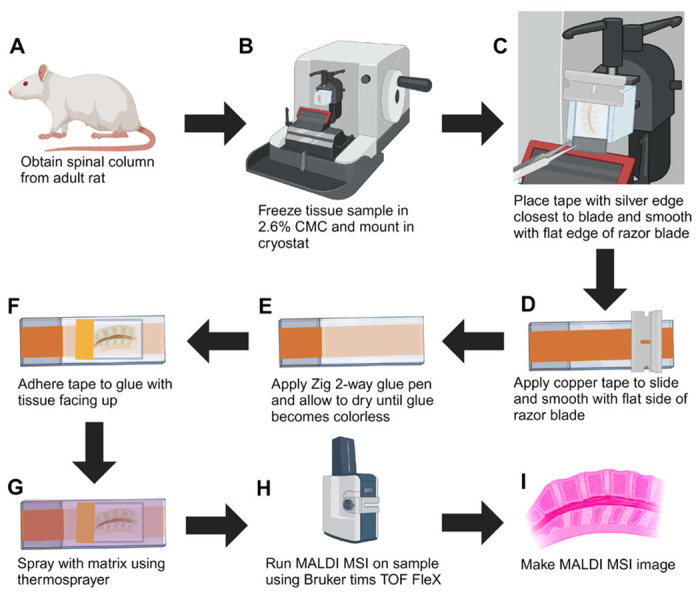
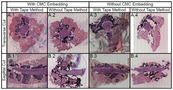
Technologies assessing the lipidomics, genomics, epigenomics, transcriptomics, and proteomics of tissue samples at single-cell resolution have deepened our understanding of physiology and pathophysiology at an unprecedented level of detail. However, the study of single-cell spatial metabolomics in undecalcified bones faces several significant challenges, such as the fragility of bone which often requires decalcification or fixation leading to the degradation or removal of lipids and other molecules and. As such, we describe a method for performing mass spectrometry imaging on undecalcified spine that is compatible with other spatial omics measurements. In brief, we use fresh-freeze rat spines and a system of carboxyl methylcellulose embedding, cryofilm, and polytetrafluoroethylene rollers to maintain tissue integrity, while avoiding signal loss from variations in laser focus and artifacts from traditional tissue processing. This reveals various tissue types and lipidomic profiles of spinal regions at 10 μm spatial resolutions using matrix-assisted laser desorption/ionization mass spectrometry imaging. We expect this method to be adapted and applied to the analysis of spinal cord, shedding light on the mechanistic aspects of cellular heterogeneity, development, and disease pathogenesis underlying different bone-related conditions and diseases. This study furthers the methodology for high spatial metabolomics of spines, as well as adds to the collective efforts to achieve a holistic understanding of diseases via single-cell spatial multi-omics.

Keywords: Lipidomics; imaging; metabolomics; neuroscience; spine.


Conflict of interest statement

The authors declare no competing financial interest.

Figures

Figure 1.
Figure 1.
Workflow for preparing undecalcified fresh-frozen spinal column for MALDI MSI. (A) Lumbar tissue is excised from adult rats. (B) Tissue embedded in 2.6% CMC for enhanced cryosectioning. (C) The silver edge of the tape is placed over the edge of the blade. (D) A conductive copper tape is applied to the slide and adhered using a razor blade. (E) ZIG 2 way glue is applied on top the copper tape and allowed to dry until colorless. (F) The tape is adhered to the glue with the tissue on the side opposite from the glue. (G) Matrix is applied via an automated sprayer (HTX M3+) to enhance ion generation and desorption (H) MALDI MSI is performed for spatial lipidomics. (I) Data analysis is performed using a variety of software packages to create lipid profiles of major spinal regions. Figure was made using Biorender.
Figure 2.
Figure 2.
H&E stained section of undecalcified, unfixed, fresh-frozen spinal column under different conditions for both transverse and sagittal sections showing bone, bone marrow, muscle, nerves, grey matter, and white matter (SI Figure 1). In brief, we assessed the effects of CMC embedding and use of cryotape for both transverse (A) and sagittal cuts (B). In general, we found that CMC embedding and the use of the cryotape preserved the tissue adequately (left column). Cryotape was required for preserving the vertebra for both transverse (A.1 and A.3) and sagittal (B.1 and B.3) sections, while CMC embedding preserved the muscle tissue surrounding the spinal cord for both transverse (A.2 and A.4) and sagittal (B.2 and B.4) sections.
Figure 3.
Figure 3.
Use of a PTFE roller resulted in fewer air bubbles and, thus, fewer imaging artifacts. (A) H&E stain of a sagittal, undecalcifled fresh-frozen spinal column embedded in CMC adhered to the slide using forceps without direct tissue contact. The presence of air bubbles between tape and glue is indicated by dark gray shadows. (B) An H&E stained tissue section adhered using a PTFE roller prior to heat fixation. (C) An H&E stained tissue section adhered using a PTFE roller after heat fixation, resulting in significant tissue degradation. (D) Example ion image of a sagittal spinal cord that was adhered using forceps as opposed to a roller (E).
Figure 4.
Figure 4.
Average mass spectrum for each region within the spinal column (n = 3) with lines pointing to the respective region. The averaged mass spectrum is shown for each region (left) and enlarged spectra covering the lipid region (right).
Figure 5.
Figure 5.
The major components of the spinal column can be visualized using MALDI MSI. such as intervertebral discs and blood vessels (A.2, B.2, m/z 725.5557, lipid [SM(34:1;O2)+Na]+), white matter (A.3, B.3, m/z 746.6041, lipid [PC(O-34:1)+H]+), bone marrow (A.4, B.4, m/z 768.5857, lipid [PC(O-34:1)+Na]+), muscle (A.5, B.5, m/z 796.5243, lipid [PC(34:2)+K]+), grey matter (A.6, B.6, m/z 834.5967, lipid [PC(38:3)+Na]+), nerves (A.7, B.7, m/z 853.6529, lipid [SM(42:1;O2)+K]+).
Figure 6.
Figure 6.
MALDI MSI layered ion images of undecalcified fresh-frozen spinal columns transversely cut from three different adult rats, normalized by total ion count. (A) Spinal column cut transversely with a raster width of 30 μm. (B) Spinal column cut transversely with a raster width of 10 μm and minimal surrounding muscle included in MALDI MSI. (C) Spinal column cut transversely with a raster width of 30 μm.

References

    1. Little J. P. Chapter 22 - The Spine: Biomechanics and Subject-Specific Finite Element Models. In DHM and Posturography; Scataglini S., Paul G., Eds.; Academic Press, 2019; pp 287–293. 10.1016/B978-0-12-816713-7.00022-2. - DOI
    1. Kayalioglu G. The V, ertebral Column and Spinal Meninges. Spinal Cord 2009, 17–36. 10.1016/B978-0-12-374247-6.50007-9. - DOI
    1. Heise C.; Kayalioglu G. Chapter 13 - Spinal Cord Transmitter Substances. In The Spinal Cord; Watson C., Paxinos G., Kayalioglu G., Eds.; Academic Press: San Diego, 2009; pp 191–208. 10.1016/B978-0-12-374247-6.50017-1. - DOI
    1. Brown A. G. Organization in the Spinal Cord: The Anatomy and Physiology of Identified Neurones; Springer Science & Business Media, 2012.
    1. McDonald J. W.; Belegu V.; Becker D. Chapter 64 - Spinal Cord. In Principles of Tissue Engineering (Fourth Edition); Lanza R., Langer R., Vacanti J., Eds.; Academic Press: Boston, 2014; pp 1353–1373. 10.1016/B978-0-12-398358-9.00064-1. - DOI

Publication types