

Posterior malleolus exposure
- PMID: 37662836
- PMCID: PMC10473347
- DOI: 10.1097/OI9.0000000000000021
Posterior malleolus exposure
Abstract
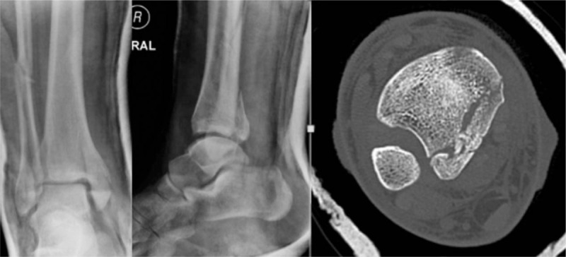
Objectives: Posterior malleolus (PM) fractures are common in rotational ankle injuries, tibial plafond fractures, and distal third tibia fractures. Surgical indications continue to evolve as we improve our understanding of ankle and syndesmotic stability. These fractures remain technically challenging with respect to both exposure and fixation. Our biomechanical cadaveric study compared posterolateral versus modified posteromedial surgical approaches to define the following: maximal surface area exposed, and maximal screw trajectory obtainable for fixation.
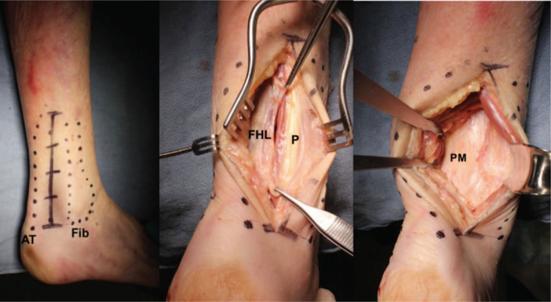
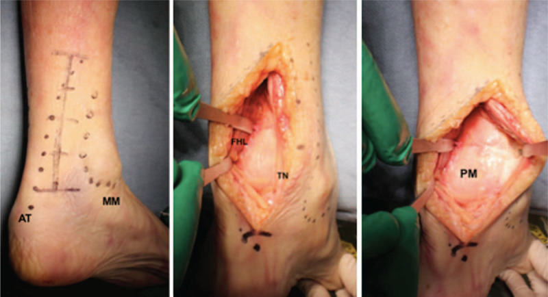
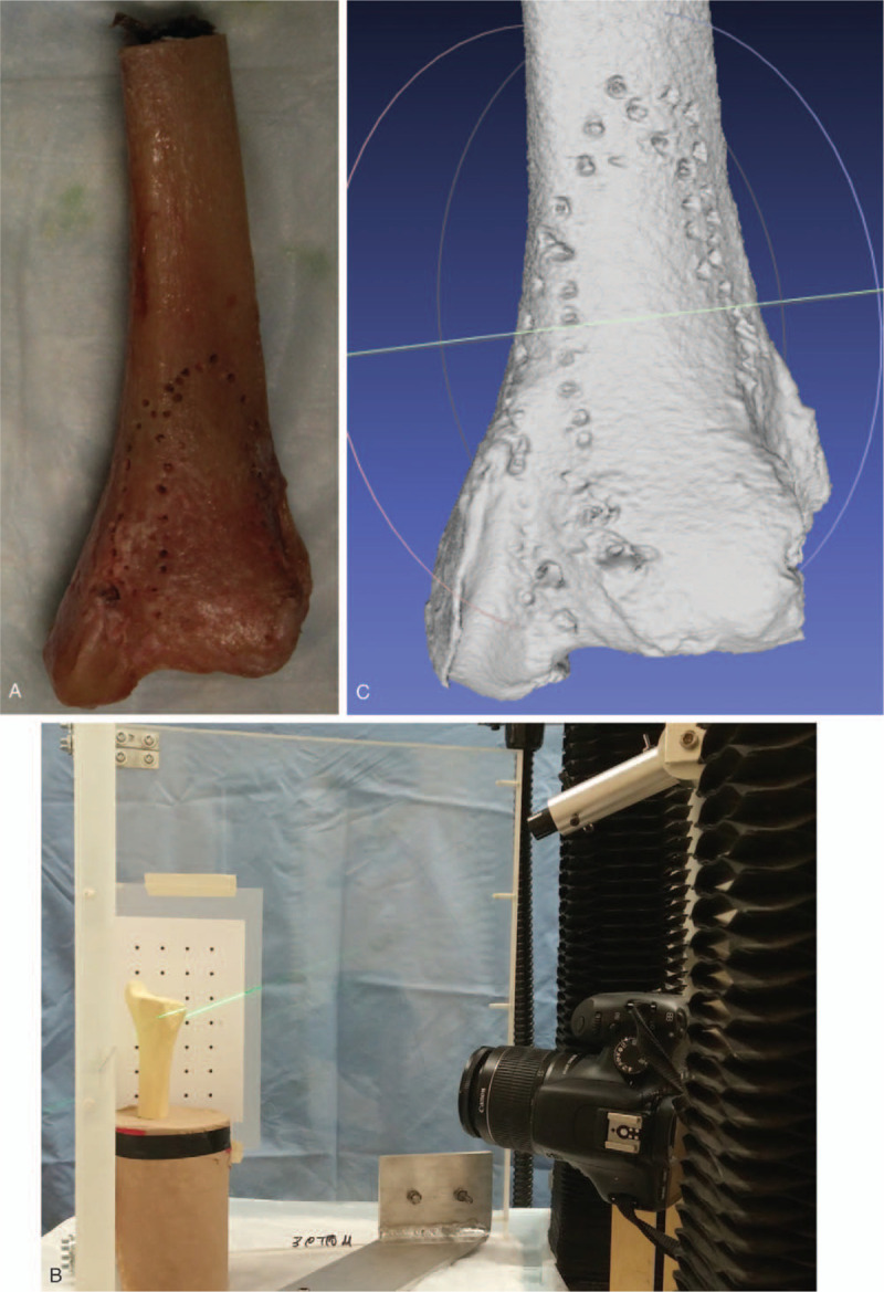
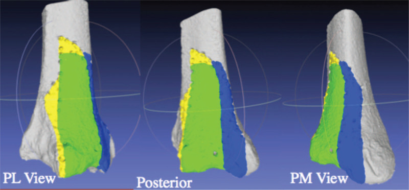
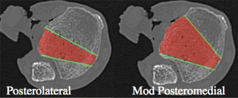
Methods: Twelve fresh-frozen cadaver limbs were thawed at room temperature. Posterolateral and modified posteromedial approaches were performed on each limb. Margins of exposure were marked. A 2.5 mm drill was advanced at the extreme medial and lateral extents of each exposure, standardized at 1 cm proximal to the joint line and perpendicular to the bone. Computed tomography (CT) scans were performed to identify the maximal trajectory. Limbs were stripped of soft tissue, and the exposed bony surface area was measured using a validated laser surface-scanning technique.
Results: The modified posteromedial approach allowed for a larger exposed surface area compared to the posterolateral exposure (median 99% vs 64%, respectively; P < .05). The modified posteromedial approach allowed for instrumentation of up to a median of 77% of the posterior distal tibia as opposed to 46% through the posterolateral approach (P < .05).
Conclusion: The modified posteromedial approach allowed for increased exposure and wider access for instrumentation of the PM when compared to the posterolateral approach. We advocate use of this approach when addressing complex PM fractures, in particular the Haraguchi type 2 fracture pattern.
Keywords: Haraguchi; ankle; approach; biomechanical; cadaver; exposure; modified posteromedial; posterior malleolus; posterolateral; screw trajectory.
Copyright © 2019 The Authors. Published by Wolters Kluwer Health, Inc. on behalf of the Orthopaedic Trauma Association.
Conflict of interest statement
The authors have no conflicts of interest to disclose.
Figures
References
-
- Gardner MJ, Streubel PN, McCormick JJ, et al. Surgeon practices regarding operative treatment of posterior malleolus fractures. Foot Ankle Int. 2011;32:385–393. - PubMed
-
- Haraguchi N, Haruyama H, Toga H, et al. Pathoanatomy of posterior malleolar fractures of the ankle. J Bone Joint Surg Am. 2006;88:1085–1092. - PubMed
-
- Assal M, Ray A, Fasel JHD, et al. A modified posteromedial approach combined with extensile anterior for the treatment of complex tibial pilon fractures (AO/OTA 43-C). J Orthop Trauma. 2014;28:e138–e145. - PubMed
-
- Irwin TA, Lien J, Kadakia AR. Posterior malleolus fracture. J Am Acad Orthop Surg. 2013;21:32–40. - PubMed
-
- Tornetta P, Ricci W, Nork S, et al. The posterolateral approach to the tibia for displaced posterior malleolar injuries. J Orthop Trauma. 2011;25:123–126. - PubMed
LinkOut - more resources
Full Text Sources