Review
. 2023 Aug 17:14:1233908.
doi: 10.3389/fimmu.2023.1233908. eCollection 2023.

Immune cells as messengers from the CNS to the periphery: the role of the meningeal lymphatic system in immune cell migration from the CNS

Affiliations
Review

Immune cells as messengers from the CNS to the periphery: the role of the meningeal lymphatic system in immune cell migration from the CNS

Collin Laaker et al. Front Immunol. .

Abstract

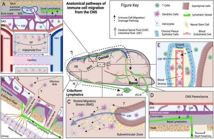
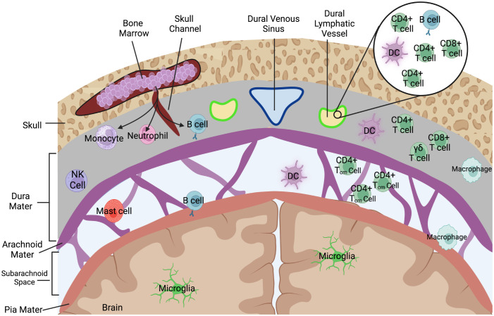
In recent decades there has been a large focus on understanding the mechanisms of peripheral immune cell infiltration into the central nervous system (CNS) in neuroinflammatory diseases. This intense research led to several immunomodulatory therapies to attempt to regulate immune cell infiltration at the blood brain barrier (BBB), the choroid plexus (ChP) epithelium, and the glial barrier. The fate of these infiltrating immune cells depends on both the neuroinflammatory environment and their type-specific interactions with innate cells of the CNS. Although the fate of the majority of tissue infiltrating immune cells is death, a percentage of these cells could become tissue resident immune cells. Additionally, key populations of immune cells can possess the ability to "drain" out of the CNS and act as messengers reporting signals from the CNS toward peripheral lymphatics. Recent data supports that the meningeal lymphatic system is involved not just in fluid homeostatic functions in the CNS but also in facilitating immune cell migration, most notably dendritic cell migration from the CNS to the meningeal borders and to the draining cervical lymph nodes. Similar to the peripheral sites, draining immune cells from the CNS during neuroinflammation have the potential to coordinate immunity in the lymph nodes and thus influence disease. Here in this review, we will evaluate evidence of immune cell drainage from the brain via the meningeal lymphatics and establish the importance of this in animal models and humans. We will discuss how targeting immune cells at sites like the meningeal lymphatics could provide a new mechanism to better provide treatment for a variety of neurological conditions.

Keywords: CNS; cervical lymph node; cribriform plate; dendritic cells; immune cell migration; meningeal lymphatics; olfactory nerves.


Conflict of interest statement

The authors declare that the research was conducted in the absence of any commercial or financial relationships that could be construed as a potential conflict of interest.

Figures

Figure 1
Figure 1
Anatomical pathways of immune cell migration from the CNS. Schematic outlining the anatomical locations of a few of the proposed pathways of immune cell migration (Dotted lines) within the brain and meningeal compartments, derived from rodent animal experiments. Common fluid movement pathways within CNS are also outlined (Blue solid lines). Each panel represents a unique anatomical site of proposed immune cell migration: (A) Dorsal dural lymphatics and Glymphatic system. Here perivascular CSF glymphatic flow is hypothesized to drive fluid transport across parenchymal space and into SAS. Immune cells are thought to utilize these perivascular pathways to reach dorsal leptomeningeal spaces (Pia, SAS, and Arachnoid). Once in the SAS, immune cells are proposed to cross the arachnoid mater and traffic to lymphatic vessels in the dural layer, potentially at “hotspots” where arachnoid barriers are more permissible such as arachnoid granulations. Evidence for this is limited however, with arachnoid granulations being primarily a human phenomenon and their relationship mostly investigated for fluid/solute flow into the superior sagittal sinus, not immune cells. (B) Olfactory Bulb-Cribriform Lymphatic axis. Olfactory nerves protrude out of the olfactory bulb through skull foramina at the cribriform plate. Along these nerves the meningeal layers have been demonstrated to be discontinuous, with potential gaps in arachnoid mater. Lymphatic vessels are closely associated with these nerve bundles and in mice they extend through the cribriform plate into the CNS-side of the skull, and potentially near these meningeal gaps. CSF and immune cells can move along this peri-nerve space into the periphery. (C) Rostral Migratory Stream (RMS). The RMS is an intra-parenchymal migration pathway for neural stem cells and evidence also suggests it facilitates the migration of dendritic cells. Starting at the subventricular zone, ISF and cells have been demonstrated to move toward the olfactory bulb along this route. Astrocytes and blood vessels populate this pathway, which are proposed to facilitate this fluid and cell movement. (D) Basal Dural Lymphatics. Here lymphatic vessels in the basal dura are proposed to facilitate immune cell egress and connect to a wider dural lymphatic system. Their close association to basal skull foramina provides a potential route to exit the skull, but similar to dorsal dural lymphatics, their communication with cells in SAS is still debated. (E) Choroid Plexus. The choroid plexus is a vascularized organ that produces CSF in the ventricles of the brain. Highly studied for its role in immune cell infiltration into the CNS, there is also limited evidence that it may participate in T cell egress from the CSF. Figure Acronyms: CSF, Cerebrospinal Fluid; ISF, Interstitial Fluid; SAS, Subarachnoid Space; OB, olfactory bulb; sCLN, Superficial cervical lymph node; dCLN, Deep cervical lymph node.
Figure 2
Figure 2
Immune cells that populate the meningeal spaces. A simplified schematic detailing the diverse repertoire of immune cells during homeostatic conditions at the dorsal meningeal spaces. The subarachnoid space is largely populated with T cells, specifically those with a CD4+ Tcm (Central memory) phenotype as well as some populations of myeloid cells including dendritic cells. Recent characterizations of skull bone marrow and dura have identified that immune cells can populate dural stroma without needing to first enter peripheral blood. Dorsal dural meningeal lymphatic vessels run alongside the superior sagittal sinus, and initial characterizations of the cellular composition of these meningeal lymphatic vessels have identified lymphocytes, DCs, and B-cells within the meningeal lymphatic lumen.

Similar articles

Cited by

References

    1. Croese T, Castellani G, Schwartz M. Immune cell compartmentalization for brain surveillance and protection. Nat Immunol (2021) 22(9):1083–92. doi: 10.1038/s41590-021-00994-2 - DOI - PubMed
    1. Castellani G, Croese T, Peralta Ramos JM, Schwartz M. Transforming the understanding of brain immunity. Science (2023) 380(6640):eabo7649. doi: 10.1126/science.abo7649 - DOI - PubMed
    1. Beuker C, Strecker JK, Rawal R, Schmidt-Pogoda A, Ruck T, Wiendl H, et al. . Immune cell infiltration into the brain after ischemic stroke in humans compared to mice and rats: a systematic review and meta-analysis. Transl Stroke Res (2021) 12(6):976–90. doi: 10.1007/s12975-021-00887-4 - DOI - PMC - PubMed
    1. Choi YH, Laaker C, Hsu M, Cismaru P, Sandor M, Fabry Z. Molecular mechanisms of neuroimmune crosstalk in the pathogenesis of stroke. Int J Mol Sci (2021) 22(17):9486. doi: 10.3390/ijms22179486 - DOI - PMC - PubMed
    1. Hurn PD, Subramanian S, Parker SM, Afentoulis ME, Kaler LJ, Vandenbark AA, et al. . T- and B-cell-deficient mice with experimental stroke have reduced lesion size and inflammation. J Cereb Blood Flow Metab (2007) 27(11):1798–805. doi: 10.1038/sj.jcbfm.9600482 - DOI - PMC - PubMed

Publication types

LinkOut - more resources