. 2023 Aug 17:14:1233128.
doi: 10.3389/fimmu.2023.1233128. eCollection 2023.

DNA and histones impair the mechanical stability and lytic susceptibility of fibrin formed by staphylocoagulase

Affiliations

DNA and histones impair the mechanical stability and lytic susceptibility of fibrin formed by staphylocoagulase

Erzsébet Komorowicz et al. Front Immunol. .

Abstract

Background: Staphylocoagulase (SCG) is a virulence factor of Staphylococcus aureus, one of the most lethal pathogens of our times. The complex of SCG with prothrombin (SCG/ProT) can clot fibrinogen, and SCG/ProT-induced fibrin and plasma clots have been described to show decreased mechanical and lytic resistance, which may contribute to septic emboli from infected cardiac vegetations. At infection sites, neutrophils can release DNA and histones, as parts of neutrophil extracellular traps (NETs), which in turn favor thrombosis, inhibit fibrinolysis and strengthen clot structure.

Objectives: To characterize the combined effects of major NET-components (DNA, histone H1 and H3) on SCG/ProT-induced clot structure, mechanical and lytic stability.

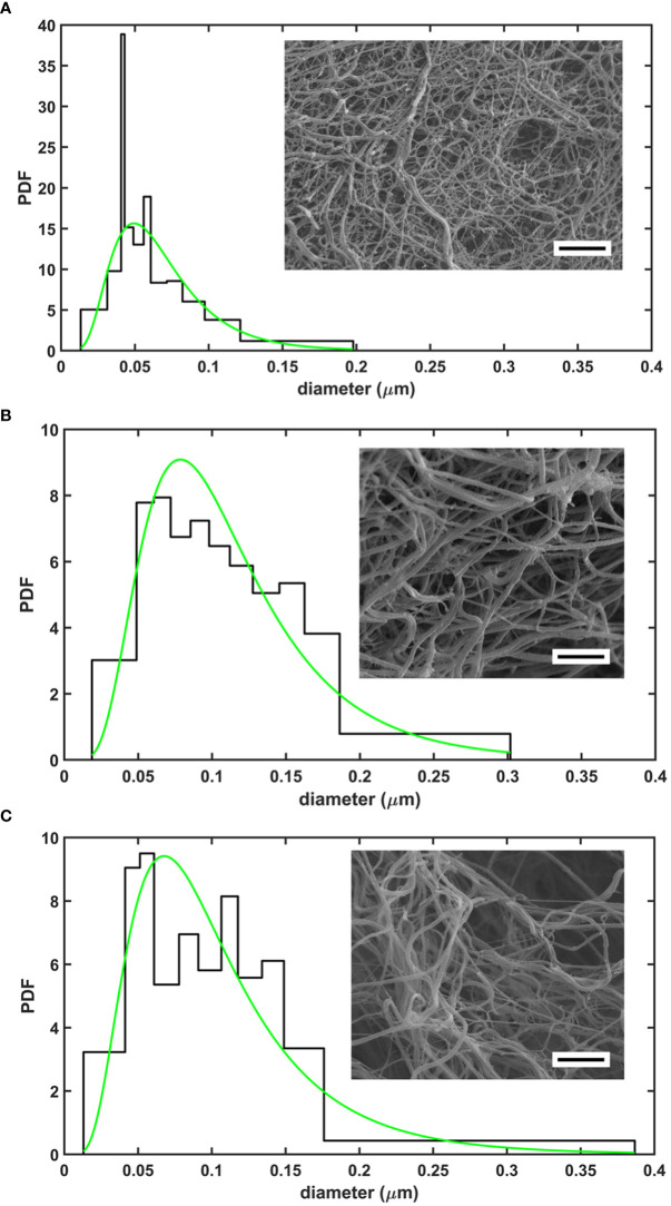
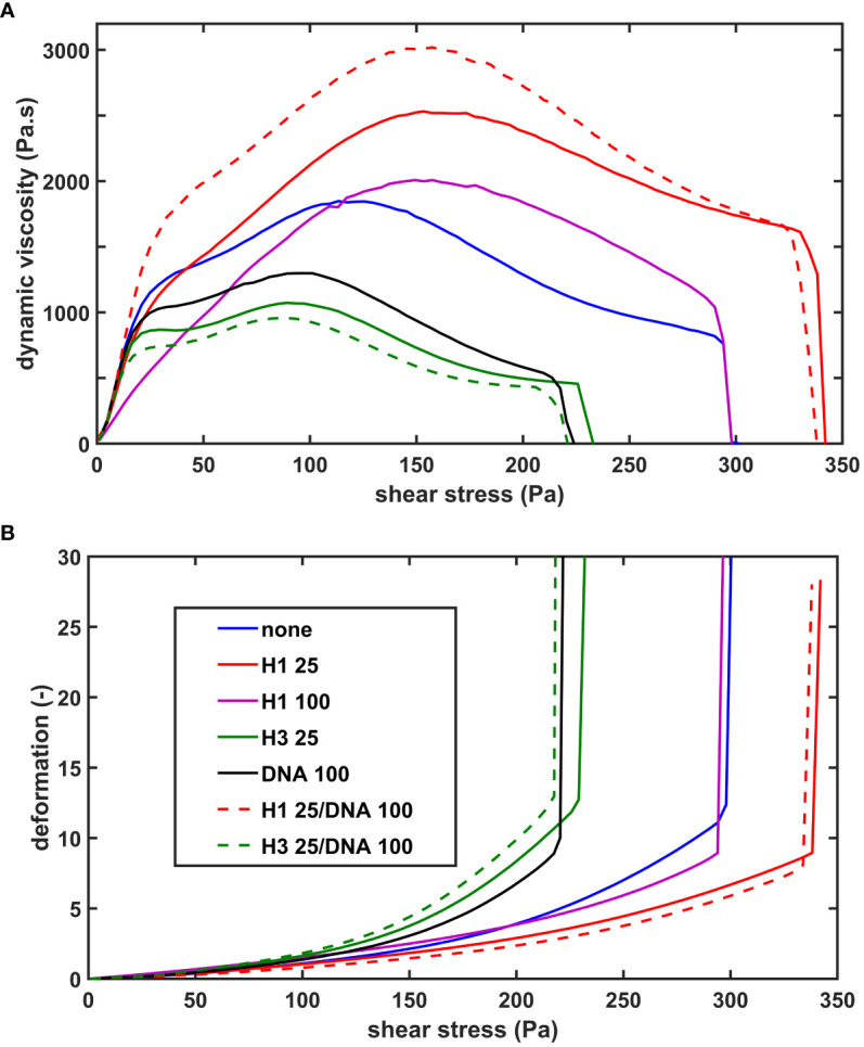
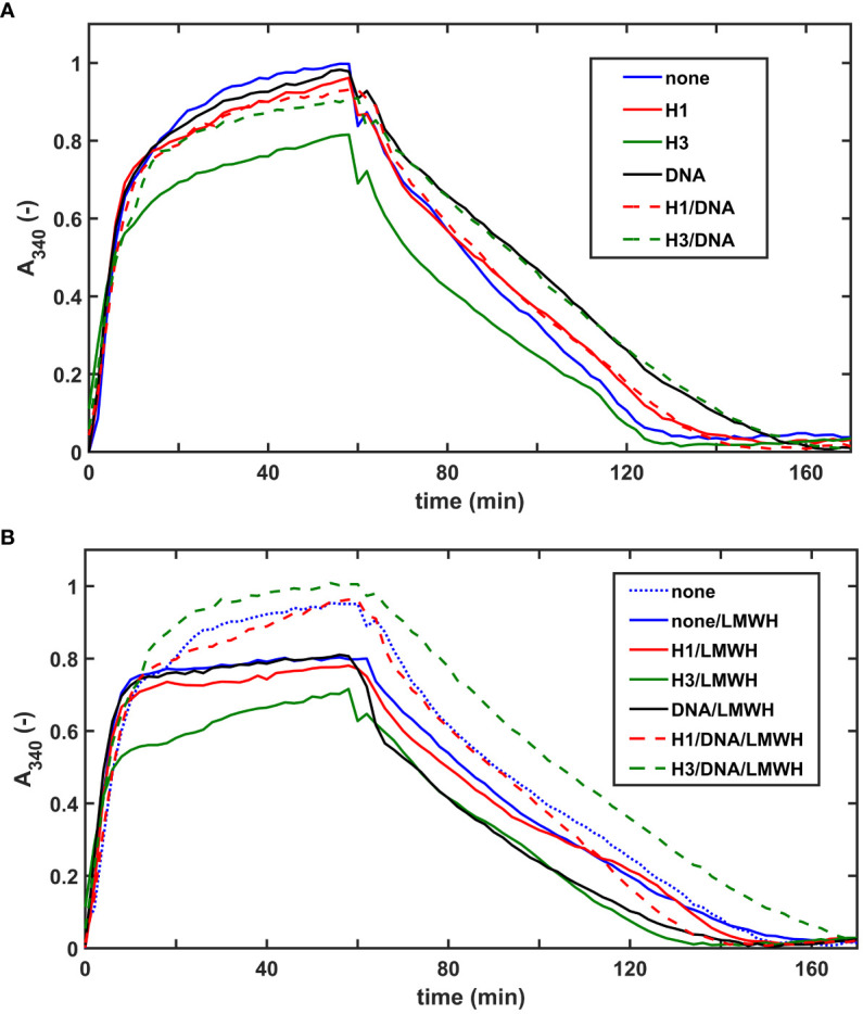
Methods: Recombinant SCG was used to clot purified fibrinogen and plasma. The kinetics of formation and lysis of fibrin and plasma clots containing H1 or core histones+/-DNA were followed by turbidimetry. Fibrin structure and mechanical stability were characterized with scanning electron microscopy, pressure-driven permeation, and oscillation rheometry.

Results: Histones and DNA favored the formation of thicker fibrin fibers and a more heterogeneous clot structure including high porosity with H1 histone, whereas low porosity with core histones and DNA. As opposed to previous observations with thrombin-induced clots, SCG/ProT-induced fibrin was not mechanically stabilized by histones. Similarly to thrombin-induced clots, the DNA-histone complexes prolonged fibrinolysis with tissue-type plasminogen activator (up to 2-fold). The anti-fibrinolytic effect of the DNA and DNA-H3 complex was observed in plasma clots too. Heparin (low molecular weight) accelerated the lysis of SCG/ProT-clots from plasma, even if DNA and histones were also present.

Conclusions: In the interplay of NETs and fibrin formed by SCG, DNA and histones promote structural heterogeneity in the clots, and fail to stabilize them against mechanical stress. The DNA-histone complexes render the SCG-fibrin more resistant to lysis and thereby less prone to embolization.

Keywords: NET; extracellular DNA; fibrin; fibrinolysis; histone; staphylocoagulase.


Conflict of interest statement

The authors declare that the research was conducted in the absence of any commercial or financial relationships that could be construed as a potential conflict of interest.

Figures

Figure 1
Figure 1
Structure of SCG/ProT-induced clots evaluated with scanning electron microscopy. Fibrin fiber distribution patterns in various clot types with a representative image are shown for fibrin formed in the absence (A), or in the presence of 75 µg/ml H1 histone (B), or 75 µg/ml H1 histone combined with 50 µg/ml DNA (C). Note the more heterogeneous structure with thicker fibers enclosing larger pores in the presence of H1 histone+/-DNA. Quantitative analysis of 4-6 images/clot type was performed as detailed in Materials and methods (scale bar is 2 µm). The probability density function (PDF) is shown for the empiric (histogram) and the fitted lognormal (green line) distributions of the diameter values. The numeric parameters of the fitted distributions are summarized in Table 1 .
Figure 2
Figure 2
The effect of NET-components on the shear resistance of SCG/ProT-induced fibrin. Fibrinogen without (none) or with supplemented NET- components (H1 histone, core histones (H3), or DNA alone or combined at the indicated concentrations in µg/ml, (see inset box) was clotted with SCG/ProT under superimposed periodic oscillatory deformation in the measurement gap of the oscillation rheometer, as detailed in Materials and Methods. After the 15 min clotting phase an increasing shear stress τ was applied to the same clot and the resulting relative deformation γ was measured. Representative flow curves of the deformation γ (B) and the corresponding computed dynamic viscosity (η) (A) are presented for the various clot types, whereas numerical data with statistics are summarized in Table 2 .
Figure 3
Figure 3
NET-components confer fibrinolytic resistance to SCG/ProT-clots in both intrinsic and extrinsic tPA-mediated models. (A) Extrinsic lysis model: fibrinogen containing 150 nM plasminogen and 75 µg/ml H1 or core histones (H3), or 50 µg/ml DNA alone, or combined as indicated was clotted in microplate wells with 8 nM SCG/ProT for 120 min and thereafter tPA (15 nM) was applied to the surface of the clot to trigger lysis. (B) Intrinsic lysis model: fibrinogen containing 15 nM plasminogen and histones and/or DNA as in panel A was clotted with 8 nM SCG/ProT in the presence of 1.3 nM tPA to induce fibrinolysis simultaneously with clotting. Ascending and descending parts of the turbidimetric curve recorded as absorbance at 340 nm wavelength indicate fibrin formation and dissolution. Presented kinetic curves are averaged from 4 parallel measurements on the same day, and numerical data shown in Table 3 ; Table S2 in the online Supplemental Material were calculated from at least 3 experiments performed on different days.
Figure 4
Figure 4
Effects of NET-components and heparins on the dissolution of pre-formed SCG/ProT-plasma clots. SCG/ProT-mediated plasma clots containing 1 µM plasminogen and 250 µg/ml H1 or core histone, or 100 µg/ml DNA, alone or combined were pre-formed in microplate wells in the absence (A) or presence (B) of 2.5 µg/ml LMWH (curve for pure plasma clot is also presented in panel B for comparison). Clot development was followed by turbidimetry as absorbance at 340 nm, and fibrinolysis was initiated at 60 min by applying 100 nM tPA on the clot surfaces. Presented kinetic curves are averaged from 4 parallel measurements on the same day, and numerical data shown in Table 4 were calculated from at least 3 such experiments performed on different days.

Similar articles

Cited by

References

    1. Liesenborghs L, Verhamme P, Vanassche T. Staphylococcus aureus, master manipulator of the human hemostatic system. J Thromb Haemost (2018) 16:441–54. doi: 10.1111/jth.13928 - DOI - PubMed
    1. Primus CP, Woldman S. Infective endocarditis remains a deadly disease – it’s bad news, especially when Staphylococci and Enterococci are involved: a call to action. J Am Heart Assoc (2022) 11:e026788. doi: 10.1161/JAHA.122.026788 - DOI - PMC - PubMed
    1. Ostregaard L, Voldstedlund M, Bruun NE, Bundgaard H, Iversen K, Kober N, et al. . Temporal changes, patient characteristics, and mortality, according to microbiological cause of infective endocarditis: a nationwide study. J Am Heart Assoc (2022) 11:025801. doi: 10.1161/JAHA.122.025801 - DOI - PMC - PubMed
    1. Garcia-Cabrera E, Fernandez-Hidalgo N, AlmIrante B, Ivanova-Georgieva R, Noureddine M, Plata A, et al. . Neurological complications of infective endocarditis. Risk factors, Outcome, and Impact of cardiac surgery: a multicenter observational study. Circulation (2013) 127:2272–84. doi: 10.1161/CIRCULATIONAHA.112.000813 - DOI - PubMed
    1. Cuervo G, Escrihuela-Vidal F, Gudiol C, Carratala J. Current challenges in the management of infective endocarditis. Front Med (2021) 8:641243. doi: 10.3389/fmed.2021.641243 - DOI - PMC - PubMed

Publication types