Review
. 2023 Aug 17:14:1225704.
doi: 10.3389/fimmu.2023.1225704. eCollection 2023.

The benefits of clustering in TNF receptor superfamily signaling

Affiliations
Review

The benefits of clustering in TNF receptor superfamily signaling

Éva S Vanamee et al. Front Immunol. .

Abstract

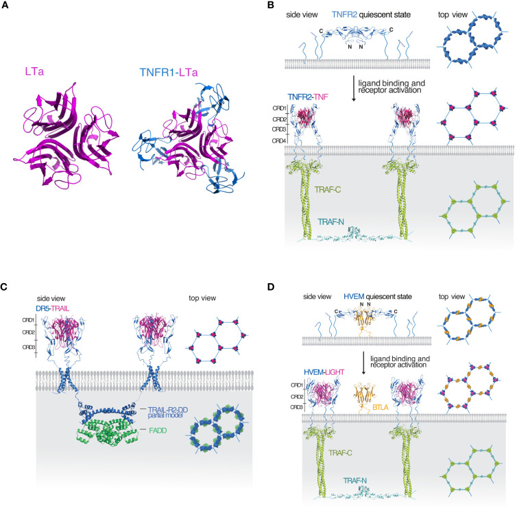
The tumor necrosis factor (TNF) receptor superfamily is a structurally and functionally related group of cell surface receptors that play crucial roles in various cellular processes, including apoptosis, cell survival, and immune regulation. This review paper synthesizes key findings from recent studies, highlighting the importance of clustering in TNF receptor superfamily signaling. We discuss the underlying molecular mechanisms of signaling, the functional consequences of receptor clustering, and potential therapeutic implications of targeting surface structures of receptor complexes.

Keywords: TNF receptor superfamily (TNFRSF); TNF signaling; TNFR agonism and antagonism; receptor clustering; signal amplification.


Conflict of interest statement

The authors declare that the research was conducted in the absence of any commercial or financial relationships that could be construed as a potential conflict of interest.

Figures

Figure 1
Figure 1
Illustration of the mechanism of signaling in the TNF receptor superfamily (A) Molecular representations of trimeric lymphotoxin (LTa) shown in magenta (top view) and the LTa (magenta)-TNF receptor 1 (TNFR1, blue) complex (top view). (B) A TRAF-interacting TNFRSF receptor represented by a model of the TNF/TNFR2/TRAF2 signaling complex. In the quiescent state (top panel), the receptor antiparallel dimers (blue) are arranged in a hexagonal lattice. TNF (magenta) binding breaks up the dimer interface and activated TNFR2 trimers recruit TRAF2 (green) resulting in the dimerization of the TRAF2 N-terminal RING domains (cyan) and activation of further downstream events. The hexagonal lattice of the downstream components mirrors the hexagonal lattice of the receptors. (C) Death receptor 5 (DR5, blue) in complex with its ligand TRAIL also forms a hexagonal cluster. After DR5 activation, TRAIL-R2-DD (blue) dimerizes and recruits a FADD dimer (green) also forming a hexagonal lattice. (D) For receptors like HVEM (blue) that are unable to dimerize on their own, hexagonal lattice formation is aided and controlled by the dimeric IgSF member, BTLA (orange). Upon binding of the ligand (magenta) the activated receptors recruit a TRAF homolog resulting in RING dimerization and activation of further downstream events. The program PyMOL was used for creating all molecular representations (3).
Figure 2
Figure 2
Illustration of signal amplification in hexagonal clusters. (A) Signal amplification represented by the e/n ratio is calculated for two examples of regular tiled hexagons with different geometries and plotted against n. It is higher in a hexagonal lattice that grows equally in both direction in the plane (shown in red) over a lattice tiled in only one direction (yellow). The red shaded area illustrates that 90-95% of the maximum signal amplification can be achieved with a cluster of 100-400 receptors in agreement with experimental data on the average size of receptor nano clusters in cells.
Figure 3
Figure 3
Illustration of simulated signal amplitudes with soluble or membrane-bound ligands. (A) Representation of a honeycomb receptor cluster with 50% occupancy. Activated receptors are represented as dark blue nodes, inactive receptors shown as light blue nodes. Edges between active nodes are shown in dark blue while all other edges are colored light blue. (B) In cell-to-cell interactions, clustered ligands attached to the membrane allow the simultaneous activation of clustered receptors with the same geometry, resulting in maximum signaling efficiency. (C) The amplitude is calculated as the ratio of active/inactive edges for 50% and 95% occupancies. High occupancy, illustrating membrane-bound ligand activation results in a sharp, high amplitude signal, while lower occupancy by soluble ligands results in a low amplitude signal spread out over time.
Figure 4
Figure 4
The size and rigidity of the stem region plays a role in receptor activation by the ligand. (A) The stem regions of several TNF receptors are listed. Soluble TNF can more readily activate TNFR1 and LTβR with short stem regions than TNFR2 that has a long stem sequence. Similarly, TRAIL can more readily activate DR4 with a short stem than DR5 that has a longer stem. (B) In the TNFR activation model the stem regions shown in light blue are exposed and may directly interact with the ligand or play an otherwise important role in receptor activation. The structures ligand bound complexes are shown in 50% transparency to indicate the final state after conformational change.
Figure 5
Figure 5
Antibody targeting strategies of TNF receptors (A) Agonist antibodies stabilize the hexagonal signaling complex TNF (magenta)-TNFR2 (blue) complexes are arranged on the cell surface in a hexagonal lattice. After receptor activation downstream TRAF signaling partners (shown in green and cyan) are recruited with matching hexagonal geometry. Agonist antibodies (shown in yellow and orange) stabilize the receptor clusters and improve signaling. (B) Antagonist antibodies stabilize the quiescent state and block the activation of receptors. Receptor dimers (blue) in the quiescent state are arranged in a hexagonal lattice on the cell surface. Antagonist antibodies shown in purple and violet lock in the ligand-free state and block ligand binding, receptor activation and the recruitment of downstream signaling partners.

Similar articles

Cited by

References

    1. Aggarwal BB. Signalling pathways of the TNF superfamily: a double-edged sword. Nat Rev Immunol (2003) 3(9):745–56. doi: 10.1038/nri1184 - DOI - PubMed
    1. Bodmer JL, Schneider P, Tschopp J. The molecular architecture of the TNF superfamily. Trends Biochem Sci (2002) 27(1):19–26. doi: 10.1016/S0968-0004(01)01995-8 - DOI - PubMed
    1. The PyMOL molecular graphics system, version 2.5, Schrödinger, LLC.
    1. Vanamee ES, Faustman DL. Structural principles of tumor necrosis factor superfamily signaling. Sci Signal (2018) 11(511). doi: 10.1126/scisignal.aao4910 - DOI - PubMed
    1. Zapata JM, Perez-Chacon G, Carr-Baena P, Martinez-Forero I, Azpilikueta A, Otano I, et al. . CD137 (4-1BB) signalosome: complexity is a matter of TRAFs. Front Immunol (2018) 9:2618. doi: 10.3389/fimmu.2018.02618 - DOI - PMC - PubMed

Substances

LinkOut - more resources