. 2023 Aug 6:33:738-748.
doi: 10.1016/j.omtn.2023.08.001. eCollection 2023 Sep 12.

Leverage of nuclease-deficient CasX for preventing pathological angiogenesis

Affiliations

Leverage of nuclease-deficient CasX for preventing pathological angiogenesis

Haote Han et al. Mol Ther Nucleic Acids. .

Abstract

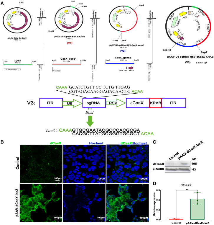
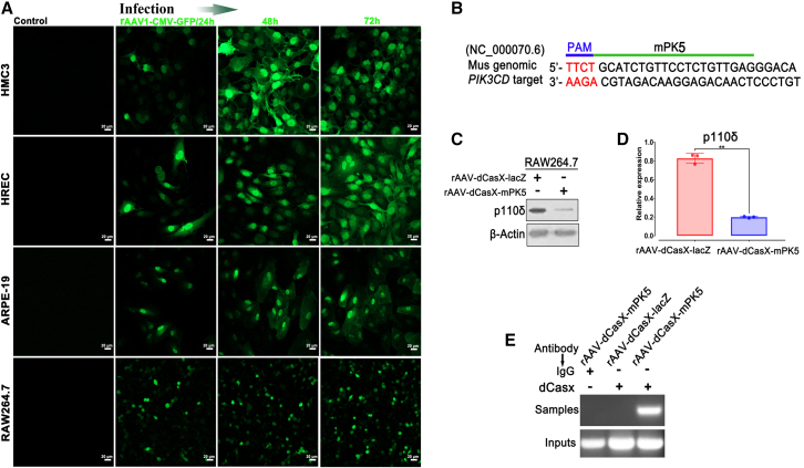
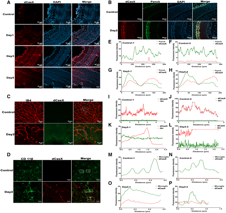
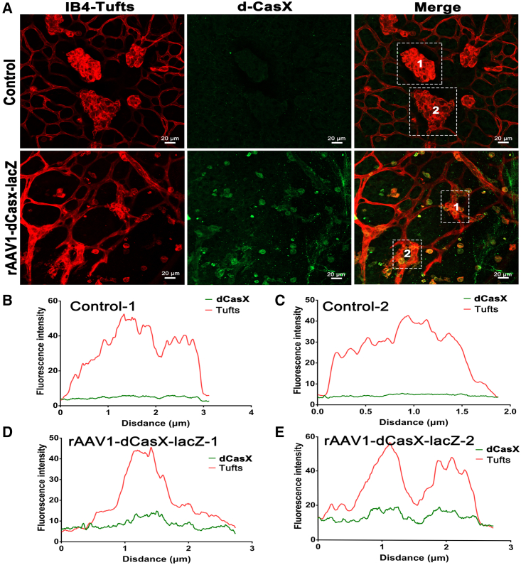
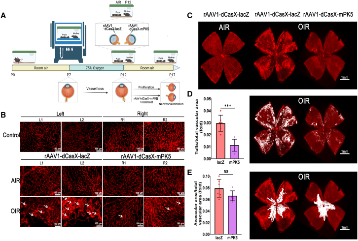
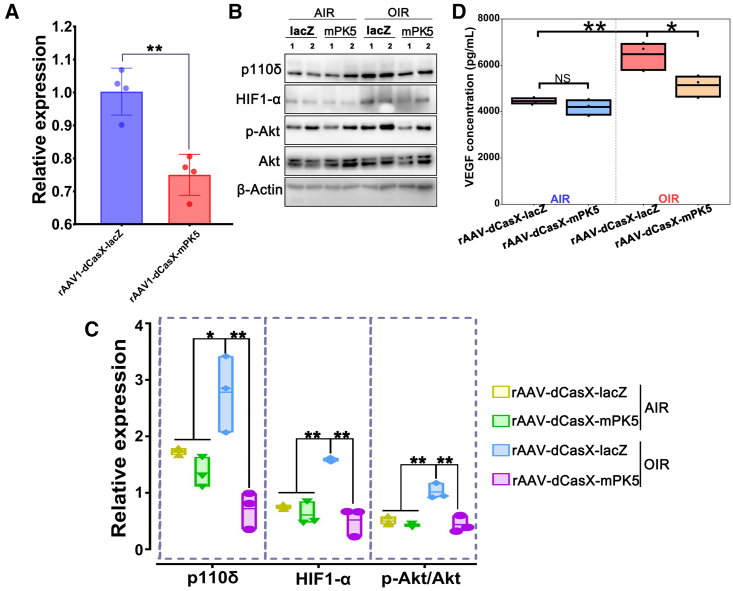
Gene editing with a CRISPR/Cas system is a novel potential strategy for treating human diseases. Pharmacological inhibition of phosphoinositide 3-kinase (PI3K) δ suppresses retinal angiogenesis in a mouse model of oxygen-induced retinopathy. Here we show that an innovative system of adeno-associated virus (AAV)-mediated CRISPR/nuclease-deficient (d)CasX fused with the Krueppel-associated box (KRAB) domain is leveraged to block (81.2% ± 6.5%) in vitro expression of p110δ, the catalytic subunit of PI3Kδ, encoded by Pik3cd. This CRISPR/dCasX-KRAB (4, 269 bp) system is small enough to be fit into a single AAV vector. We then document that recombinant AAV serotype (rAAV)1 efficiently transduces vascular endothelial cells from pathologic retinal vessels, which show high expression of p110δ; furthermore, we demonstrate that blockade of retinal p110δ expression by intravitreally injected rAAV1-CRISPR/dCasX-KRAB targeting the Pik3cd promoter prevents (32.1% ± 5.3%) retinal p110δ expression as well as pathological retinal angiogenesis in a mouse model of oxygen-induced retinopathy. These data establish a strong foundation for treating pathological angiogenesis by AAV-mediated CRISPR interference with p110δ expression.

Keywords: CRISPR/dCasX-KRAB; MT: RNA/DNA editing; PI3Kδ; Pik3cd; angiogenesis; oxygen-induced retinopathy; rAAV1.


Conflict of interest statement

All the authors declare that they have no conflicts of interest with the contents of this article.

Figures

None
Graphical abstract
Figure 1
Figure 1
Establishment of an AAV vector expressing CRISPR/dCasX-KRAB (A) Schematic diagram of constructing an AAV-CRISPR/dCasX-KRAB (dCasX) vector. The pAAV-U6-sgRNA/RSV-SpCas9 (V1) vector was originated from pAAV-RSV-SpCas9 (Addgene, 85450). The pAAV-U6-sgRNA/RSV-CasX_gene1 (V2) was derived from V1 by replacement of the Cas9 with CasX_gene1. The pAAV-U6-sgRNA/RSV-dCasX (V3) was originated from V2. ITR, inverted terminal repeat; U6, a promoter of polymerase III; RSV, a promoter of RSV. (B) HEK293T cells were transfected with the plasmid of pAAV-dCasX/sgRNA-lacZ. 48 h later, the transfected cells were immunostained with a primary antibody against CasX. Green signals indicate dCasX expression. Scale bar, 100 μm. (C). Lysates of transfected HEK293T cells were subjected to Western blot analysis using indicated antibodies. A representative of at least three independent experiments is shown. (D) The bar graphs are mean ± SD of three independent experiments. The data was analyzed using one-way ANOVA followed by the Tukey honest significant difference post hoc test. ∗∗p < 0.01.
Figure 2
Figure 2
CRISPR/dCasX-KRAB-mediated blockage of p110δ expression in vitro (A) HRECs, ARPE-19 cells and RAW264.7 were infected with rAAV1 virus carrying dCasX/sgRNA-lacZ. After 24, 48, and 72 h, the infected cells were immunostained with a primary antibody against dCasX. Green signals indicate dCasX expression. Scale bar, 20 μm. (B) Graphical schematic of a mouse Pik3cd locus. mPK5 sgRNA was originated from the Pik3cd promoter. The PAM is marked in blue. (C and D) Raw264.7 cells were infected with rAAV1-dCasX/sgRNA-lacZ and dCasX/sgRNA-mPK5, respectively. The transfected cells were subjected to western blotting analyses 48 h later (C and D). The bar graphs are mean ± SD of three independent experiments. The data was analyzed using one-way ANOVA followed by the Tukey honest significant difference post hoc test. ∗∗p < 0.01. (E) DNA samples from a ChIP assay with RAW264.7 cells, which were infected with rAAV1-dCasX/sgRNA-lacZ and dCasX/sgRNA-mPK5, respectively, were subjected to PCR and agarose gel electrophoresis analysis. Non-immune IgG served a negative control.
Figure 3
Figure 3
rAAV1-delivered dCasX expression in vivo (A and B) Normal P12 mice were injected intravitreally with rAAV1 carrying dCasX/sgRNA-lacZ (1μL, 1.62 × 10E13 vg/mL). After injection for 1, 3, and 5 days, frozen eyeball sections were stained with a primary antibody against dCasX. Red signals indicate dCasX expression, and green indicate Panck expression. Scale bar, 20 μm. (C and D) Whole-mount retinas from the intravitreally injected in mice were stained with IB4 (red) and antibodies against dCasX and CD11β (green) and then with a fluorescent-labeled secondary antibody. Images were obtained in the immunofluorescence confocal microscope. Scale bars, 10 or 20 μm. (E–P) Co-localization analysis of dCasX expression in vivo was performed by ImageJ. Two regions were selected for each image.
Figure 4
Figure 4
rAAV1 infects pathological vascular ECs with high efficiency (A) P12 mice that had been exposed to 75% oxygen for 5 days were injected intravitreally with rAAV1-dCasX/sgRNA-lacZ (1μL, 1.62 × 10E13 vg/mL). Whole-mount retinas from the P17 mice in a model of OIR were stained with IB4 (red) and a primary antibody against dCasX and then a fluorescent-labeled secondary antibody. Scale bar, 20 μm. (B–E) Co-localization analysis of dCasx expression with tufts in vivo was performed by ImageJ. Two regions were selected for each image.
Figure 5
Figure 5
CRISPR/dCasX-KRAB-mediated blockade of p110δ expression suppresses pathological angiogenesis in a mouse model of OIR (A) Schematic graph of the OIR model. Litters of P12 mice that had been exposed to 75% oxygen for 5 days were injected intravitreally with rAAV1 (1 μL, 1.62 × 10E13 vg/mL) carrying dCasX/sgRNA-lacZ or dCasX/sgRNA-mPK5. (B) The whole-mount retinas from the injected P17 mice were stained with IB4. Arrows indicate tufts. AIR indicates mice that were kept in room air without exposure to 70% oxygen. L, left eye of the mice; R, right eye of the mice. Scale bar, 100 μm. (C–E) Quantification of tufts and avascular areas as described previously. The bar graphs are mean ± SD (n = 6). The data were analyzed using one-way ANOVA followed by the Tukey honest significant difference post hoc test. ∗∗∗p < 0.01. NS, not significant.
Figure 6
Figure 6
dCasX-mediated p110δ attenuation suppresses Akt activation and VEGF production in the mouse model of OIR (A and B) Retinal mRNA and proteins from P17 mice (n = 6) subjected to qPCR (A) and western blotting analyses with indicated antibodies (B), respectively. (C) The intensity of bands. The bar graphs are mean ± SD of three independent experiments. The data were analyzed using one-way ANOVA followed by the Tukey honest significant difference (HSD) post hoc test. ∗p < 0.05, ∗∗p < 0.01. (D) Clarified vitreous (5 μL) from the P17 mice with or without experiencing OIR was subjected to ELISA analysis by following the instructions of a Quantikine Mouse VEGF ELISA Kit. The bar graphs are mean ± SD of six mice. The data were analyzed using one-way ANOVA followed by the Tukey HSD post hoc test. NS, not significant. ∗p < 0.05; ∗∗p < 0.01.

Similar articles

Cited by

References

    1. Šuleková M., Fitzgerald K.T. Can the Thought of Teilhard de Chardin Carry Us Past Current Contentious Discussions of Gene Editing Technologies? Camb. Q. Healthc. Ethics. 2019;28:62–75. - PubMed
    1. Gasiunas G., Sinkunas T., Siksnys V. Molecular mechanisms of CRISPR-mediated microbial immunity. Cell. Mol. Life Sci. 2014;71:449–465. - PMC - PubMed
    1. Wagner J.C., Platt R.J., Goldfless S.J., Zhang F., Niles J.C. Efficient CRISPR/Cas9-mediated genome editing in P. falciparum. Nat. Methods. 2014;11:915–918. - PMC - PubMed
    1. Gu P., Yang Q., Chen B., Bie Y.N., Gu W. Genetically blocking HPD via CRISPR/Cas9 protects against lethal liver injury in a pig model of tyrosinemia type I. Mol. Ther. Methods Clin. Dev. 2021;21:530–547. - PMC - PubMed
    1. Shinmyo Y., Tanaka S., Tsunoda S., Hosomichi K., Tajima A., Kawasaki H. CRISPR/Cas9-mediated gene knockout in the mouse brain using in utero electroporation. Sci. Rep. 2016;6:20611. - PMC - PubMed