

Asymptomatic congenital ductus arteriosus aneurysm in a newborn: Case by approach
- PMID: 37663558
- PMCID: PMC10473969
- DOI: 10.1016/j.radcr.2023.08.050
Asymptomatic congenital ductus arteriosus aneurysm in a newborn: Case by approach
Abstract
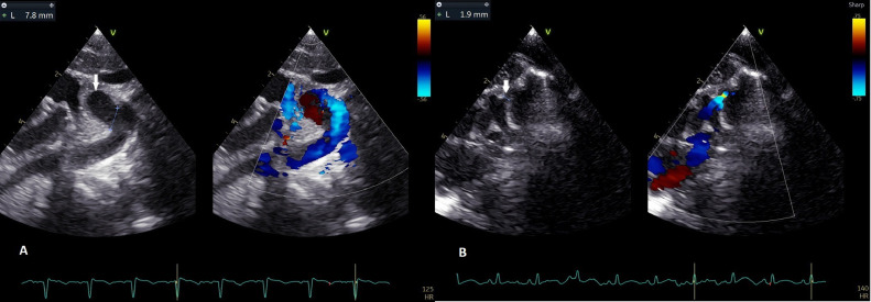
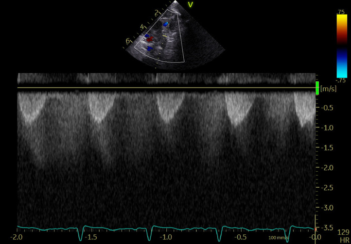
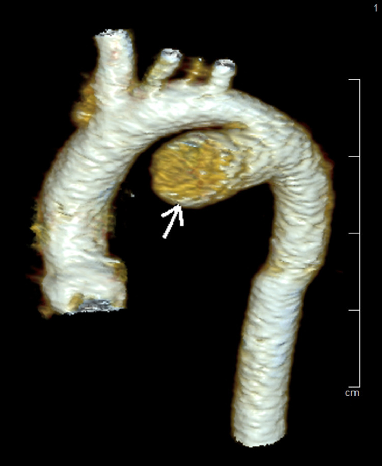
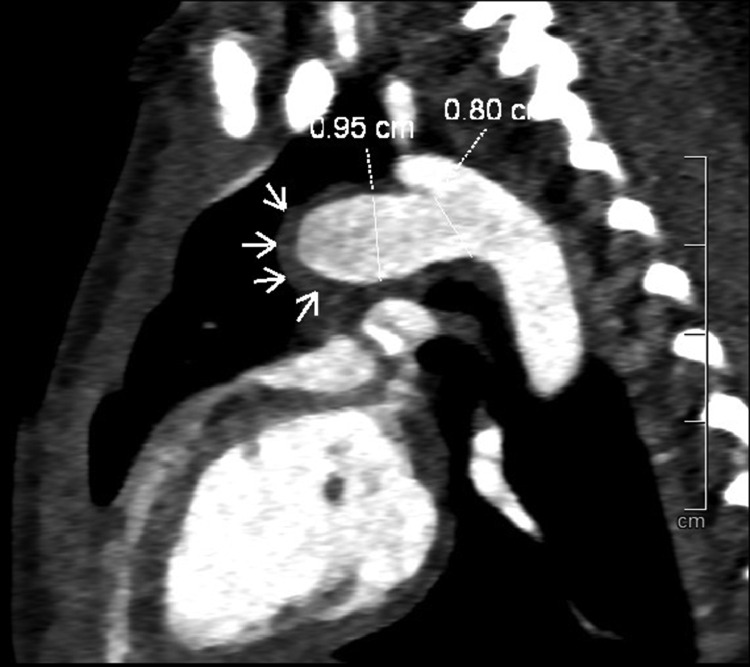
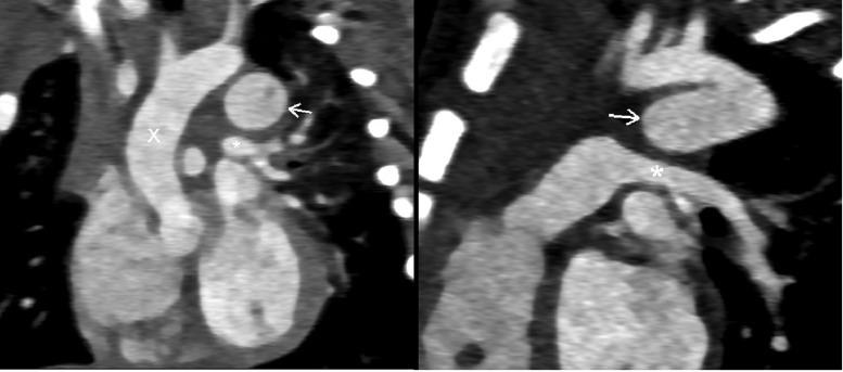
A ductus arteriosus aneurysm is a rare congenital lesion with a localized saccular or tubular dilation of the ductus arteriosus. This lesion usually appears in all ages. Some case reports suggest the most common age of diagnosis is less than 2 months. We reported a case of an asymptomatic ductus arteriosus aneurysm in neonates. Echocardiography at 2 days of age revealed a tubular dilation of the ductus arteriosus connected to the pulmonary artery. Computed tomography angiogram showed a ductus arteriosus aneurysm with thrombus at the pulmonary end. It resolved spontaneously in the six months of life without serious complications.
Keywords: Computed tomography angiogram; Ductus arteriosus aneurysm; Echocardiography; Patent ductus arteriosus; Thrombosis.
© 2023 The Authors.
Figures
Similar articles
-
Aneurysm of the ductus arteriosus: a congenital lesion.Am J Perinatol. 1998;15(12):653-9. doi: 10.1055/s-2007-999298. Am J Perinatol. 1998. PMID: 10333391 Clinical Trial.
-
Prenatal sonographic diagnosis of congenital ductus arteriosus aneurysm: a case report.J Med Assoc Thai. 2005 Apr;88(4):541-4. J Med Assoc Thai. 2005. PMID: 16146263
-
Ductus arteriosus aneurysm with left pulmonary artery obstruction.Echocardiography. 2021 Jul;38(7):1128-1130. doi: 10.1111/echo.15070. Epub 2021 May 16. Echocardiography. 2021. PMID: 33998041
-
Ductus arteriosus aneurysm: Case report and review of the literature.Arch Pediatr. 2018 May;25(4):283-285. doi: 10.1016/j.arcped.2018.03.005. Epub 2018 Apr 12. Arch Pediatr. 2018. PMID: 29656824 Review.
-
Aneurysm of the ductus arteriosus in the neonate: three case reports with a review of the literature.Pediatr Cardiol. 1992 Oct;13(4):222-6. doi: 10.1007/BF00838780. Pediatr Cardiol. 1992. PMID: 1518741 Review.
Cited by
-
Acute thrombosis of ductus arteriosus aneurysm causing bilateral pulmonary artery occlusion in a neonate.J Cardiothorac Surg. 2024 Dec 23;19(1):680. doi: 10.1186/s13019-024-03179-8. J Cardiothorac Surg. 2024. PMID: 39716226 Free PMC article.
-
Ductus arteriosus aneurysm in neonates.BMC Cardiovasc Disord. 2025 Jul 4;25(1):464. doi: 10.1186/s12872-025-04925-z. BMC Cardiovasc Disord. 2025. PMID: 40615799 Free PMC article.
References
-
- Amato J.J., Cardarelli M.G., Bierman F.Z. Aneurysm of the arterial duct—a case report and review of the literature. Cardiol Young. 1994;4(1):87–89. doi: 10.1017/S1047951100010969. - DOI
Publication types
LinkOut - more resources
Full Text Sources
