. 2023 Aug 12;9(3):100341.
doi: 10.1016/j.jve.2023.100341. eCollection 2023 Sep.

Sulforaphane prevents the reactivation of HIV-1 by suppressing NFκB signaling

Affiliations

Sulforaphane prevents the reactivation of HIV-1 by suppressing NFκB signaling

Imran Jamal et al. J Virus Erad. .

Abstract

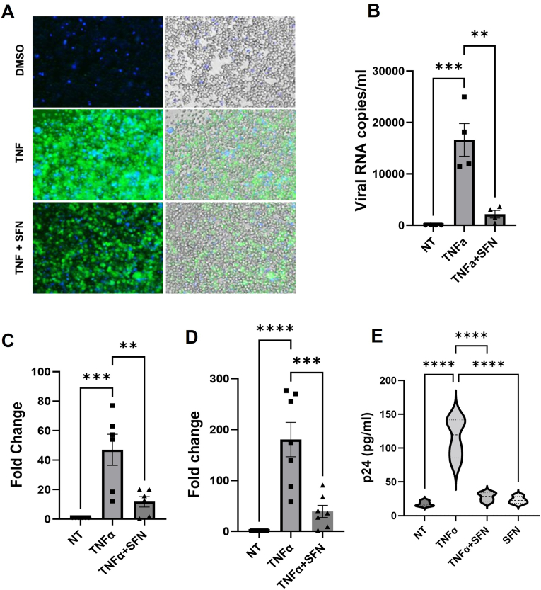
Despite more than 20 years of combination antiretroviral therapy (cART), complete eradication of HIV remains a daunting task. While cART has been very effective in limiting new cycles of infection and keeping viral load below detectable levels with partial restoration of immune functions, it cannot provide a cure. Evidently, the interruption of cART leads to a quick rebound of the viral load within a few weeks. These consistent observations have revealed HIV ability to persist as an undetectable latent reservoir in a variety of tissues that remain insensitive to antiretroviral therapies. The 'Block-and-Lock' approach to drive latent cells into deep latency has emerged as a viable strategy to achieve a functional cure. It entails the development of latency-promoting agents with anti-HIV functions. Recent reports have suggested sulforaphane (SFN), an inducer of NRF-2 (nuclear erythroid 2-related factor 2)-mediated antioxidative signaling, to possess anti-HIV properties by restricting HIV replication at the early stages. However, the effect of SFN on the expression of integrated provirus remains unexplored. We have hypothesized that SFN may promote latency and prevent reactivation. Our results indicate that SFN can render latently infected monocytes and CD4+ T cells resistant to reactivation. SFN treatments antagonized the effects of known latency reactivating agents, tumor necrosis pactor (TNF-α), and phorbol 12-myristate 13-acetate (PMA), and caused a significant reduction in HIV transcription, viral RNA copies, and p24 levels. Furthermore, this block of reactivation was found to be mediated by SFN-induced NRF-2 signaling that specifically decreased the activation of NFκB signaling and thus restricted the HIV-1 promoter (5'LTR) activity. Overall, our study provides compelling evidence to highlight the latency-promoting potential of SFN which could be used in the 'Block-and-Lock' approach to achieve an HIV cure.


Conflict of interest statement

The authors declare that they have no known competing financial interests or personal relationships that could have appeared to influence the work reported in this paper.

Figures

Fig. 1
Fig. 1
SFN treatment prevents the reactivation of HIV-1 in THP89 cells (latently infected monocyte cell line). A) HIV-1 latently infected monocyte THP89GFP cells were treated with a media-supplemented vehicle only (DMSO) or with TNF-alpha 10 ng/ml in the presence or absence of SFN (10μM). Twenty-four hours after treatment, Cells were observed by fluorescent microscopy at 20× magnification. B) HIV-1 RNA copies released in the supernatant after treatment with TNF-α (10 ng/ml) and SFN (10 μM). Cell supernatants were collected, and genomic HIV-1 RNA was purified using a viral RNA purification kit. RT-qPCR assay was used to quantify HIV-1 RNA copy numbers in the supernatant. C) and D) Quantitative RT-PCR assays were performed on cellular RNA collected 24h post-treatment using primers specific for HIV-1 initiation (C) and elongation (D) transcripts. Expression is represented as fold change over the non-treated sample. GAPDH was used as internal control to normalize the amount of HIV-1 RNA (and cDNA) in each reaction. E) ELISAs were performed to measure HIV-1 p24 level in the supernatant of THP89-GFP cells after treatment with 10 ng/ml TNF-α and 10 μM SFN for 24h. Data is represented as violin plots. Each column shows mean value with error bars indicating the standard deviation. ** indicates p<0.01 and *** indicates p<0.001 by one-way ANOVA, Dunnett's Multiple Comparisons testing.
Fig. 2
Fig. 2
SFN treatment protects the reactivation of HIV-1 in U1 cells (latently infected monocyte cell line). A) Cell supernatants were collected from the U1 HIV-1 latently infected monocyte U1 line which were treated with media-supplemented vehicle only (NT) or with TNF-alpha 10 ng/ml in the presence or absence of SFN (10μM) for 24 h. Genomic viral RNA copy numbers were quantified by RT-qPCR. B) and C) Quantitative RT-PCR assays were performed on cellular RNA collected 24h post-treatments using primers specific for HIV initiation (B) and elongation (C) transcripts. Expression is represented as fold change over the non-treated sample. GAPDH was used as internal control to normalize the amount of RNA in each reaction. D) ELISAs were performed to measure HIV-1 p24 level in supernatants after respective treatment for 24h. Data is represented as violin plots. Each column shows the mean value with error bars indicating the standard deviation. ** indicates p<0.01 and *** indicates p<0.001 by one-way ANOVA, Dunnett's Multiple Comparisons testing.
Fig. 3
Fig. 3
SFN treatment prevents the reactivation of HIV-1 in J89GFP and ACH2 cells (latently infected T cell lines). A) and B) Cellular RNA was collected from HIV-1 latently infected J89GFP cells 24h after the respective treatments and quantitative RT-PCR assays were performed using primers specific for HIV initiation (A) and elongation (B) transcripts. C) ELISAs were performed to measure HIV-1 p24 level in supernatants after respective treatments for 24h. D) J89GFP cells were reactivated with PMA (15 nM) in the presence or absence of SFN (10μM) for 24 h and HIV-1 p24 level in supernatants was quantified by ELISA. E) ACH2 cells were reactivated with PMA (15 nM) in the presence or absence of SFN (10μM) for 24h.Viral RNA copy numbers in supernatant were quantified by RT-qPCR assay. Each column shows the mean value with error bars indicating the standard deviation. ** indicates p<0.01 and *** indicates p<0.001 by one-way ANOVA, Dunnett's Multiple Comparisons testing.
Fig. 4
Fig. 4
Sulforaphane has no cytotoxic effects on uninfected as well as latently infected cells. Cellular viability was determined after treating latently infected and uninfected parental cell lines with TNF-α (10 ng/ml) and SFN (10 μM). After 24h of treatment latently infected THP89 (A), J89 (B) and uninfected Jurkat (C) and THP1 (D) cells were stained with annexin V. Data is presented as mean ± standard deviation.
Fig. 5
Fig. 5
Sulforaphane activates NRF-2 signaling pathway and modulates activation of NFkB in HIV-1 latently infected cells. HIV-1 latently infected THP89GFP and J89GFP cells were treated with TNF-alpha (10 ng/ml) in the presence or absence of SFN (10uΜ). Proteins from whole cell lysates were resolved by SDS-PAGE and identified by western blotting using antibodies with the indicated specificities (A, D, G). Densitometric analysis was performed using the NIH Image analysis software on the NRF2-2, p-NFkB bands and normalized to the values of the corresponding GAPDH bands and expressed as fold change (B, C, E, F, H, and I). Each column indicates the mean value with error bars indicating the standard deviation. * Indicates p<0.05 by one-way ANOVA, Dunnett's Multiple Comparisons testing.
Fig. 6
Fig. 6
Sulforaphane treatment attenuated TNF-alpha-induced induction of NF-kB and HIV-1 LTR activity. HIV-1 latently infected THP89GFP and J89GFP cells were stimulated with TNF-alpha (10 ng/ml) in the presence or absence of SFN (10mΜ) for 15 min. Whole cell extracts from THP89 (A) and J89GFP cells (B) after respective treatments were analyzed for the binding of NFkB to its consensus DNA binding element using the TransAM NFkB p65 Kit. C) HIV-1 LTR promoter activity was measured in luciferase reporter TZMBL cells using the Promega Bright-Glo Luciferase Assay kit. Each column shows the mean value with error bars indicating the standard deviation. *** indicates p<0.001 and **** indicates p<0.0001 by one-way ANOVA, Dunnett's Multiple Comparisons testing.

References

    1. Kumar A., Abbas W., Herbein G. HIV-1 latency in monocytes/macrophages. Viruses. 2014;6:1837–1860. doi: 10.3390/v6041837. - DOI - PMC - PubMed
    1. Honeycutt J.B., Thayer W.O., Baker C.E., et al. HIV persistence in tissue macrophages of humanized myeloid-only mice during antiretroviral therapy. Nat Med. 2017;23:638–643. doi: 10.1038/nm.4319. - DOI - PMC - PubMed
    1. Kandathil A.J., Sugawara S., Balagopal A. Are T cells the only HIV-1 reservoir? Retrovirology. 2016;13:86. doi: 10.1186/s12977-016-0323-4. - DOI - PMC - PubMed
    1. Wallet C., de Rovere M, van Assche J, et al. Microglial cells: the main HIV-1 reservoir in the brain. Front Cell Infect Microbiol. 2019;9:362. doi: 10.3389/fcimb.2019.00362. - DOI - PMC - PubMed
    1. Yukl S.A., et al. The distribution of HIV DNA and RNA in cell subsets differs in gut and blood of HIV-positive patients on ART: implications for viral persistence. J Infect Dis. 2013;208:1212–1220. doi: 10.1093/infdis/jit308. - DOI - PMC - PubMed