

Recent advances in Fe-based bioresorbable stents: Materials design and biosafety
- PMID: 37663617
- PMCID: PMC10474570
- DOI: 10.1016/j.bioactmat.2023.07.024
Recent advances in Fe-based bioresorbable stents: Materials design and biosafety
Abstract
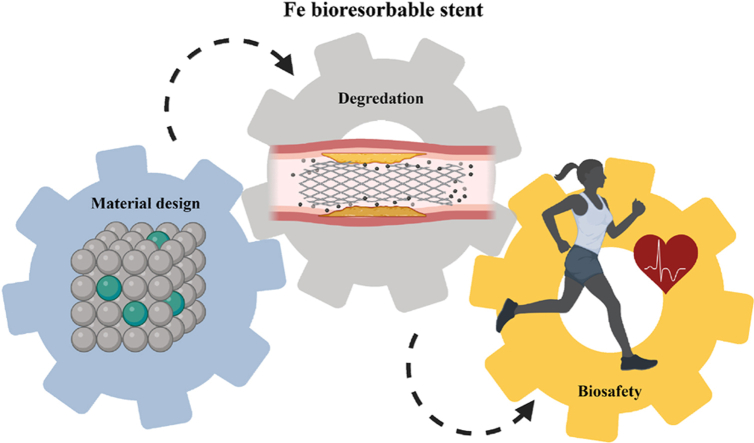
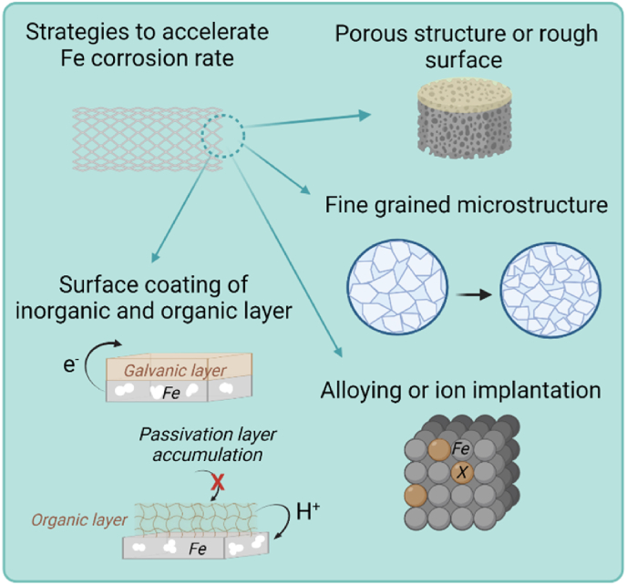
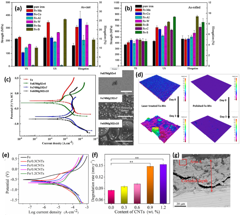
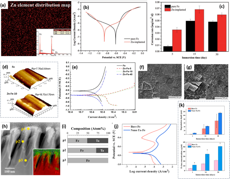
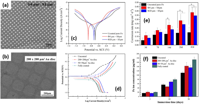
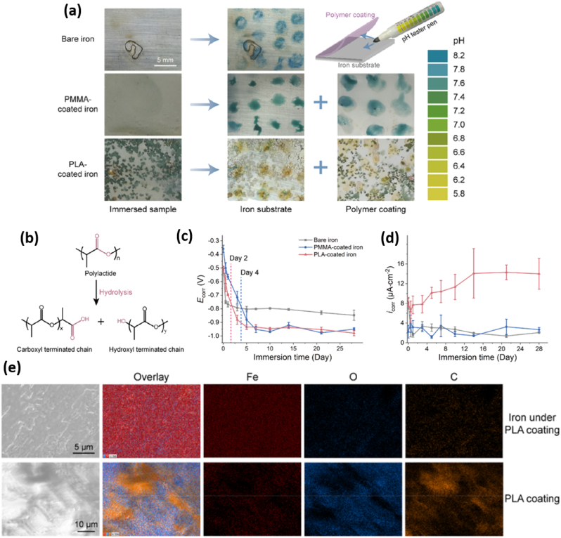
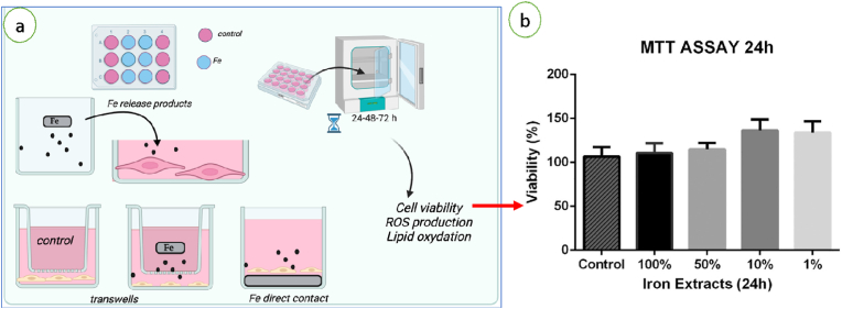
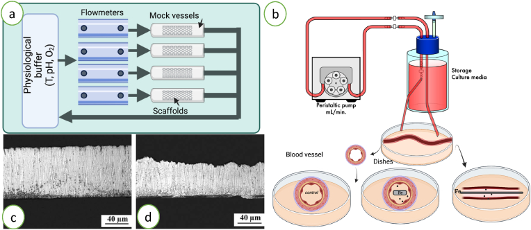
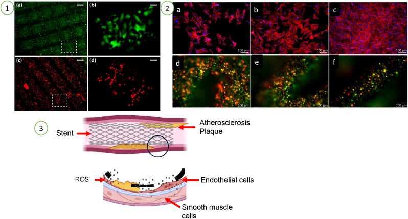
Fe-based materials have received more and more interests in recent years as candidates to fabricate bioresorbable stents due to their appropriate mechanical properties and biocompatibility. However, the low degradation rate of Fe is a serious limitation for such application. To overcome this critical issue, many efforts have been devoted to accelerate the corrosion rate of Fe-based stents, through the structural and surface modification of Fe matrix. As stents are implantable devices, the released corrosion products (Fe2+ ions) in vessels may alter the metabolism, by generating reactive oxygen species (ROS), which might in turn impact the biosafety of Fe-based stents. These considerations emphasize the importance of combining knowledge in both materials and biological science for the development of efficient and safe Fe-based stents, although there are still only limited numbers of reviews regarding this interdisciplinary field. This review aims to provide a concise overview of the main strategies developed so far to design Fe-based stents with accelerated degradation, highlighting the fundamental mechanisms of corrosion and the methods to study them as well as the reported approaches to accelerate the corrosion rates. These approaches will be divided into four main sections, focusing on (i) increased active surface areas, (ii) tailored microstructures, (iii) creation of galvanic reactions (by alloying, ion implantation or surface coating of noble metals) and (iv) decreased local pH induced by degradable surface organic layers. Recent advances in the evaluation of the in vitro biocompatibility of the final materials and ongoing in vivo tests are also provided.
© 2023 The Authors.
Conflict of interest statement
Potential conflict of interest exists: No conflict of interest exists.
Figures
Similar articles
-
The enhancement of mechanical properties and uniform degradation of electrodeposited Fe-Zn alloys by multilayered design for biodegradable stent applications.Acta Biomater. 2023 Apr 15;161:309-323. doi: 10.1016/j.actbio.2023.02.029. Epub 2023 Feb 27. Acta Biomater. 2023. PMID: 36858165
-
Cytotoxicity of corrosion products of degradable Fe-based stents: relevance of pH and insoluble products.Colloids Surf B Biointerfaces. 2015 Apr 1;128:480-488. doi: 10.1016/j.colsurfb.2015.02.047. Epub 2015 Mar 6. Colloids Surf B Biointerfaces. 2015. PMID: 25797480
-
A manufacturing and annealing protocol to develop a cold-sprayed Fe-316L stainless steel biodegradable stenting material.Acta Biomater. 2019 Nov;99:479-494. doi: 10.1016/j.actbio.2019.08.034. Epub 2019 Aug 23. Acta Biomater. 2019. PMID: 31449928
-
Recent advances and directions in the development of bioresorbable metallic cardiovascular stents: Insights from recent human and in vivo studies.Acta Biomater. 2021 Jun;127:1-23. doi: 10.1016/j.actbio.2021.03.058. Epub 2021 Apr 3. Acta Biomater. 2021. PMID: 33823325 Review.
-
Modifications on porous absorbable Fe-based scaffolds for bone applications: A review from corrosion and biocompatibility viewpoints.J Biomed Mater Res B Appl Biomater. 2022 Jan;110(1):18-44. doi: 10.1002/jbm.b.34893. Epub 2021 Jun 15. J Biomed Mater Res B Appl Biomater. 2022. PMID: 34132457 Review.
Cited by
-
Application of biodegradable implants in pediatric orthopedics: shifting from absorbable polymers to biodegradable metals.Bioact Mater. 2025 Apr 10;50:189-214. doi: 10.1016/j.bioactmat.2025.04.001. eCollection 2025 Aug. Bioact Mater. 2025. PMID: 40256329 Free PMC article.
-
Bioresorbable Materials for Wound Management.Biomimetics (Basel). 2025 Feb 12;10(2):108. doi: 10.3390/biomimetics10020108. Biomimetics (Basel). 2025. PMID: 39997131 Free PMC article. Review.
-
Semiconducting polymer dots for multifunctional integrated nanomedicine carriers.Mater Today Bio. 2024 Mar 24;26:101028. doi: 10.1016/j.mtbio.2024.101028. eCollection 2024 Jun. Mater Today Bio. 2024. PMID: 38590985 Free PMC article. Review.
-
Biodegradable high-nitrogen iron alloy anastomotic staples: In vitro and in vivo studies.Bioact Mater. 2024 Jun 5;40:34-46. doi: 10.1016/j.bioactmat.2024.06.005. eCollection 2024 Oct. Bioact Mater. 2024. PMID: 38910967 Free PMC article.
-
Polydiolcitrate-MoS2 Composite for 3D Printing Radio-Opaque, Bioresorbable Vascular Scaffolds.ACS Appl Mater Interfaces. 2024 Aug 28;16(34):45422-45432. doi: 10.1021/acsami.4c07364. Epub 2024 Aug 5. ACS Appl Mater Interfaces. 2024. PMID: 39102678 Free PMC article.
References
-
- Mendis S., Puska P., Norrving B. World Health Organization. World Stroke Organization; Copenhagen: 2011. Global Atlas on Cardiovascular Disease Prevention and Control.
-
- Vaduganathan M., Mensah G.A., Turco J.V., Fuster V., Roth G.A. The global burden of cardiovascular diseases and risk: a compass for future health. J. Am. Coll. Cardiol. 2022;80(25):2361–2371. - PubMed
-
- Zain M.A., Jamil R.T., Siddiqui W.J. StatPearls. StatPearls Publishing; Treasure Island (FL): 2022. Neointimal hyperplasia.
-
- Ruan Y., Yang R. Relationship between stent fracture and stent thrombosis. Nat. Rev. Cardiol. 2020;17(1) 64-64. - PubMed
-
- Guillory R.J., Bowen P.K., Hopkins S.P., Shearier E.R., Earley E.J., Gillette A.A., Aghion E., Bocks M., Drelich J.W., Goldman J. Corrosion characteristics dictate the long-term inflammatory profile of degradable zinc arterial implants. ACS Biomater. Sci. Eng. 2016;2(12):2355–2364. - PubMed
Publication types
LinkOut - more resources
Full Text Sources
Research Materials
