Review
. 2023 Aug 27:31:368-394.
doi: 10.1016/j.bioactmat.2023.08.016. eCollection 2024 Jan.

Research advances of nanomaterials for the acceleration of fracture healing

Affiliations
Review

Research advances of nanomaterials for the acceleration of fracture healing

Mo Zhang et al. Bioact Mater. .

Abstract

The bone fracture cases have been increasing yearly, accompanied by the increased number of patients experiencing non-union or delayed union after their bone fracture. Although clinical materials facilitate fracture healing (e.g., metallic and composite materials), they cannot fulfill the requirements due to the slow degradation rate, limited osteogenic activity, inadequate osseointegration ability, and suboptimal mechanical properties. Since early 2000, nanomaterials successfully mimic the nanoscale features of bones and offer unique properties, receiving extensive attention. This paper reviews the achievements of nanomaterials in treating bone fracture (e.g., the intrinsic properties of nanomaterials, nanomaterials for bone defect filling, and nanoscale drug delivery systems in treating fracture delayed union). Furthermore, we discuss the perspectives on the challenges and future directions of developing nanomaterials to accelerate fracture healing.

Keywords: Bone defect; Bone regeneration; Delayed union; Fracture healing; Nanomaterials; Non-union.


Conflict of interest statement

The authors declare no conflict of interest.

Figures

Image 1
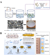
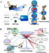
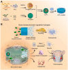
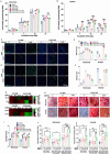
Graphical abstract
Fig. 1
Fig. 1
Illustration of a typical fracture healing process, (A) biological events, and (B) cellular activities at different stages.
Fig. 2
Fig. 2
Timeline of the representative breakthrough of nanomaterials for fracture healing in recent years.
Fig. 3
Fig. 3
Schematic illustration of rapid cutaneous wound repair and tissue regeneration using MSN-Ceria as a ROS-scavenging tissue adhesive [39]. Copyright 2017, Elsevier Ltd.
Fig. 4
Fig. 4
(A) Schematic demonstration of the metabolic activity assessment of collagen gel-seeded stem cells. (B) The total metabolic activity per initial number of cells was measured at different time points after 2 h exposure to 200 of μM H2O2. (C) The inverse of the cell shape index of stem cells from various conditions on the fourth-day post-2 h –H2O2 exposure. (D) Representative fluorescence images of the CellROX® Green in the stem cells for the intracellular oxidative stress analysis; *p < 0.05. (E) The mean gray value of CellROX® Green per cell was quantified using ImageJ software. (F) The activity in stem cells after 2 h exposure to culture medium or medium containing 200 μM of H2O2; *p < 0.05. (G) Schematic illustration of CAM assay. The eggshell was opened to expose CAM after 12 days of fertilization. (H) Quantifying the total number of blood vessels shown in the cross-sectional area of CAM where the sample was implanted; *p < 0.05. (I) Quantifying the percentage of blood vessel number in the respective range of cross-sectional area concerning the total blood vessel number in the CAM; *p < 0.05 [40]. Copyright 2019, Elsevier Ltd.
Fig. 5
Fig. 5
(A) Fabrication of Ti scaffolds by curling after deposition of a PPy-PDA-HA film on a 2D Ti mesh. (B) Preparation of the PPy-PDA-HA film via the LBL-PED process. Each PPy-PDA or HA layer was deposited in situ on a sublayer during this process. (C) The PPy-PDA-HA-coated porous scaffold was electroactive, cell affinitive, antioxidative, and osteoinductive [43]. Copyright 2019, Wiley-VCH Verlag GmbH.
Fig. 6
Fig. 6
(A) Schematic illustration of the fabrication and osteoimmunomodulatory and antibacterial properties of the nAg/HNTs/GelMA hybrid hydrogel [52]. Copyright 2019, Elsevier B.V. (B) Schematic preparation of bioresorbable dual-purpose porous microspheres with nanosilver and apatite mounted to regenerate the infected bone defect [53]. Copyright 2018, WILEY-VCH Verlag GmbH.
Fig. 7
Fig. 7
(A) Schematic illustration of the possible reaction setting of Cu-NGp. (B) Amount of silicate, Ca2+, and Cu2+ ions released from the nano glass paste for one day. (C) MSC viability of serially diluted extracts (1 original ∼ 1/256) of Cu-NGp and NGp in standard culture media, measured by CCK assay on day two (n = 6). (D–E) Expression of osteogenic genes (RUNX2 and OCN) with extracts of 1/16 and 1/32 on days five and ten by qRT-PCR assay (n = 3). An osteogenic medium (OM) was used for the osteogenesis control. (F) ALP activity measured up to ten days (n = 3). (G–H) Cellular mineralization was detected by ARS staining and the quantification on day 14 (n = 6). Cu-NGp osteopromotive at similar levels to Cu-free NGp. Statistical significance was noted when compared to control or OM (#) or compared between groups (*) at p < 0.05. (I–J) Ex ovo CAM modeling illustrated the analyses of samples implanted for four days for the neo-vessel formation in terms of total length, size, and junction. Data normalized to control (Ctrl) showed a significant difference compared with Ctrl (#), and between groups (*), at p < 0.05, for n = 8–11 [58]: copyright 2020, Elsevier Ltd.
Fig. 8
Fig. 8
(A) AgNPs increase mMSCs proliferation. (B) MTT assay was performed to test the effect of AgNPs on mMSC viability. **p<0.01 (n = 3). (C) AgNPs promote mMSC osteoblast differentiation. mMSCs were cultured in OM with various concentrations of AgNPs for 21 days; cells were then stained with Alizarin Red S (n = 3). (D) AgNPs activate TGF-β and BMP signaling pathways in fracture sites during healing. Regions highlighted in broken lines were enlarged and shown underneath. Original magnification × 40 (n = 6). Abbreviations: m: marrow; nc: new callus [63]. Copyright 2015, Elsevier Inc.
Fig. 9
Fig. 9
(A) Schematic illustration of a possible mechanism that HAp MPs with different hierarchical structures could influence the regulation of osteogenic differentiation of stem cells. (B) The expression of essential proteins in the ERK, JNK, and p38 signaling pathways in mBMSCs cultured with the three HAp MPs for 14 days. (C) The fold change of the gray value level relative to GAPDH, n = 2 [65]; copyright 2020, The Royal Society of Chemistry.
Fig. 10
Fig. 10
(A) Schematic illustration of a possible mechanism that ZS/HA/Col scaffolds with 10 wt% ZS activated the p38 signaling pathway in monocytes. (B) Micro-CT imaging of rat critical-sized cranial defects implanted with ZS/HAp/Col scaffolds containing different proportions of zinc silicate at 8 and 12 weeks postsurgery. No platforms were embedded in the blank group. The newly formed bone is indicated in yellow; the media is shown in gray; scale bar: 5 mm. (C) Bone volume fraction. (D) Residual material volume fraction. (E) Trabecular thickness (Tb. Th). (F) Trabecular number (Tb. N). (G–H) Statistical comparisons of BMP-2 (G) and Osterix (H) transcription levels in vivo two weeks after implantation. αp < 0.05 vs. 0 ZS/HA/Col scaffolds; βp < 0.05 vs. 5 ZS/HAp/Col scaffolds; γp < 0.05 vs. 10 ZS/HA/Col scaffolds; δp < 0.05 vs 15 ZS/HA/Col scaffolds [70]. Copyright 2020, American Chemical Society.
Fig. 11
Fig. 11
(A) Representative images of TRAP staining of BMMs administrated with 30 ng mL−1 M-CSF and 50 ng/mL RANKL for two days (top panel) and four days (bottom panel) in the presence of different concentrations of fullerenol; scale bar: 200 μm. (B) Fullerenol reduces the number of TRAP-positive cells with (pre-osteoclast) formation. (C) The number of TRAP-positive multinuclear cells (nuclei >3) in each well (96-well plate) was counted. For B and C, error bars are mean ± SD of triplicate experiments, *p < 0.05, ***p < 0.001, a significant difference compared to 0 μM fullerenol groups [77]. Copyright 2017, The Royal Society of Chemistry. (D) Multinucleated (≥ three nuclei) TRAP-positive cells were quantified, averaging three wells ± Stdev and representing three independent experiments. Cells were administered with RANKL (15 ng/mL) for 16 h and harvested for RNA analysis of (E) NFATC1 or (F) RANK by qRT-PCR expressed as fold change relative to the untreated control Avg. ± StDev. *p < 0.05, **p < 0.01, and ***p < 0.005 relative to the RANKL-treated Students test (n = 3) [78]. Copyright 2018, Elsevier Ltd.
Fig. 12
Fig. 12
Illustration of the inhibitory effect of IONPs on RANKL-induced osteoclastogenesis. (A) After systematic administration, IONPs are detained in the bone marrow by phagocytosis of BMMs. IONPs could inhibit RANKL-induced osteoclastogenesis in the early differentiation and late maturation stages while not affecting cell proliferation and fusion. (B) Molecular mechanism of the inhibitory effect on RANKL-induced osteoclastogenesis [80]. Copyright 2019, Elsevier Ltd.
Fig. 13
Fig. 13
(A) Schematic illustration of S + B preparation (E) scaffold; the sequential release of SDF-1 and BMP-2 from stand facilitates BMSC homing, proliferation, and osteogenic differentiation [92]. Copyright 2016, Elsevier Ltd. (B) Schematic illustration of B + V scaffold preparation; controlled dual release of low BMP-2 and VEGF synergistically affected vascularized bone regeneration [92,93]. Copyright 2017, The Royal Society of Chemistry.
Fig. 14
Fig. 14
Diagram of the pro-regenerative immune response mechanism of bone mimetic nHAp particles (BMnP) [95]. Copyright 2020, Elsevier Ltd.
Fig. 15
Fig. 15
(A) Schematic illustration of topography constructed on the HAp scaffold and the effects on osteogenic differentiation [98]. Copyright 2020, American Chemical Society. (B) Schematic illustration of the topography of the Sr-HAp scaffolds and their effects on osteogenic differentiation [99]. Copyright 2020, MDPI. (C) Schematic illustration of bone regeneration ability of 3D-printed pure HAp/TCP pure ceramic scaffolds with variable pore architectures [102]. Copyright 2020, MDPI.
Fig. 16
Fig. 16
Schematic diagram of the fabrication process for nHAC/PLGA/GO scaffolds [116]. Copyright 2018, Springer Nature.
Fig. 17
Fig. 17
(A) Levofloxacin was successfully loaded with MSNs by electrostatic attraction [126]. Copyright 2017, Science Edition. (B) Schematic illustration of antibiotic-loaded bone cement preparation. (C) Gentamicin release profiles of PMMA-based bone cement formulated with MSN, HAp, and CNT compared with antibiotic bone cement SmartSET GHV [127]. Copyright 2015, Elsevier B.V. (D) Cumulative GTMC release profiles of TNT/GTMC formulated Simplex-P bone cement with different TNTs contents. (E) The compression strength of TNTs prepared Simplex-P bone cement before and after the drug release study [128]. Copyright 2018, Elsevier Ltd.
Fig. 18
Fig. 18
(A) Cell survival was analyzed at different bFGF@MSNs concentrations. *p < 0.05 versus a 0 μg/L bFGF@MSNs concentration. (B) The proliferation of MC3T3-E1 cells cultured with or without nanoparticles (MSNs or bFGF@MSNs) in a standard medium. *p < 0.05 versus the control group, #p < 0.05 versus the MSNs group. (C) Representative images of the morphologies of MC3T3-E1 cells. Green represents the cytoskeleton, and blue represents the cell nucleus. (D) Representative images of live/dead staining of MC3T3-E1 cells. Green represents live cells, and red represents dead cells [129]. Copyright 2022, Dove Medical Press Limited.
Fig. 19
Fig. 19
(A) Bone-targeting SAA liposome for delayed fracture union [137]. Copyright 2018, Elsevier Inc. (B) Schematic diagram illustrating Asp8-liposome-icaritin. Icaritin was loaded into the bilayer lipid membrane in a liposome, and Asp8 was supplemented to target the bone surface [138]. Copyright 2017, Elsevier Inc. (C) Affinity of the BPA-liposomes to HAp. (D) Cytotoxicities of the DOX-loaded liposomes, with and without BPA, as functions of the initial drug concentrations. (E) The comparison of the cytotoxicities of the liposomes without DOX as functions of liposome concentrations. The results are the mean ± SD (n = 3) [139]. Copyright 2009, Elsevier Ltd.
Fig. 20
Fig. 20
(A) Histological specimens from calvarial defects 8, 10, and 12 weeks after implanting bone graft substitutes with hematoxylin-eosin staining. (B) Histological quantification study of new bone matrix formation at 8, 10, and 12 weeks after implanting bone graft substitutes. **p<0.0001 compared to controls and between treatment groups, respectively [148]. Copyright 2015, Dove Medical Press Limited. (C) Schematic illustration of the multifunctional BPs/MT@PLGA-ALE NSs for treating osteoporotic fracture with NIR irradiation. ALE: alendronate; BPs: black phosphorus nanosheets; MT: melatonin; OBs: osteoblasts; OCs: osteoclasts; OVX: ovariectomy [150]. Copyright 2023, Wiley-VCH GmbH.
Fig. 21
Fig. 21
(A) Schematic representation of stimuli-responsive mechanized Zr-MOFs (UiO-66-NH2) with positively charged A stalks encircled by carboxylatopillar arene (CP5) rings on the surfaces. The mechanized UiO-66-NH2 Zr-MOFs can be operated with pH variations, Ca2+ concentrations, and thermotherapy, releasing 5-Fu. (B) Controlled release profiles of the 5-Fu-loaded, CP5-capped UiO-66-NH-A can be regulated. Operation by Ca2+ addition and the release percent of 5-Fu for two days as a function of Ca2+ concentration [158]. Copyright 2021, The Royal Society of Chemistry.
Fig. 22
Fig. 22
(A) The possible formation mechanism of a 3D cellulose-HAp nanocomposite enriched with Dex-loaded metal-organic frameworks [160]. Copyright 2019, Springer Nature. (B) The DEX@Zn–Mg-MOF74/PDA composite coating preparation process and physical pictures of pristine and functionalized PEEK samples [161]. Copyright 2021, American Chemical Society.
Fig. 23
Fig. 23
Schematic diagram of VCM-NPs/Gel for osteomyelitis therapy [168]. Copyright 2020, Dove Medical Press Limited.
Fig. 24
Fig. 24
(A) The mechanisms underlying the ability of HA@SDF-1α/M2D-Exos hydrogel to accelerate fracture healing [172]. Copyright 2023, Elsevier B.V. (B) The means of HA-based natural polymer hydrogel to accelerate fracture repair [173]. Copyright 2022, American Chemical Society.
Fig. 25
Fig. 25
Schematic illustration of bone microenvironment regulative hydrogels with ROS scavenging and prolonged oxygen generation to enhance bone repair. (A) The PFC loaded in PLGA/PPS nanoparticles to form PFC@PLGA/PPS nanoparticles. Liposomes were used to co-load PFC@PLGA/PPS nanoparticles and CAT to construct CPP-L, which further encapsulated in a GelMA hydrogel and finally built up the CPP-L/GelMA intelligent response oxygen-releasing hydrogel. (B) The CPP-L/GelMA can act as a “bone microenvironment regulative hydrogel” to reverse the hypoxic microenvironment in the bone defects region to promote osteogenesis [177]. Copyright 2019, Springer Nature B.V.
Fig. 26
Fig. 26
Osteogenic differentiation of hMSCs encapsulated in oxygen-generating and osteoinductive hydrogels with different concentrations of SNPs and OMPs under normoxia and anoxia. (A–B) ALP activity normalized to the total amount of DNA of encapsulated hMSCs in pristine, OMP, SNP, or SNP/OMP constructs under (A) normoxia and (B) anoxia. (n = 3; ***p < 0.001, **p < 0.01, *p < 0.05). (C–D) Fluorescence microphotographs and (E–F) associated semiquantitative image analysis of hMSC-laden hydrogels immunohistochemically stained for OPN and OCN after being cultured in osteogenic differentiation media under normoxic or anoxic conditions. (n = 3; ****p < 0.0001, ***p < 0.001, **p < 0.01, *p < 0.05). (G) QRT-PCR analysis for three relative mRNA expression levels of late osteogenic markers OPN and OCN from hMSCs encapsulated in pristine, OMP, SNP, and SNP/OMP hydrogels after being cultured under normoxic or anoxic conditions in osteogenic differentiation media for three weeks. (n = 3). (H) Microphotographs and (I) semi-quantitative image analysis of Alizarin red stained hMSCs laden hydrogels after being cultured in osteogenic differentiation media under normoxic or anoxic conditions for 28 days. (n = 3; ***p < 0.001, **p < 0.01). The compressive modulus of hMSC-laden hydrogels in pristine OMP, SNP, and SNP/OMP hydrogels for 7 or 35 days using distinct temporal exposure to proliferation and osteogenic differentiation media under (J) normoxic or (K) anoxic conditions. Scale bar (C) = 100 μm, (H) = 25 μm. (n = 3; ****p < 0.0001, **p < 0.01, *p < 0.05) [179]. Copyright 2019, Elsevier Ltd.

References

    1. Toosi S., Behravan N., Behravan J. Nonunion fractures, mesenchymal stem cells and bone tissue engineering. J. Biomed. Mater. Res., Part A. 2018;106:2552–2562. - PubMed
    1. Chen W., Lv H., Liu S., Liu B., Zhu Y., Chen X., Yang G., Liu L., Zhang T., Wang H., Yin B., Guo J., Zhang X., Li Y., Smith D., Hu P., Sun J., Zhang Y. National incidence of traumatic fractures in China: a retrospective survey of 512 187 individuals. Lancet Global Health. 2017;5:807–817. - PubMed
    1. Lv H., Chen W., Zhang T., Hou Z., Yang G., Zhu Y., Wang H., Yin B., Guo J., Liu L., Hu P., Liu S., Liu B., Sun J., Li S., Zhang X., Li Y., Zhang Y. Traumatic fractures in China from 2012 to 2014: a national survey of 512,187 individuals. Osteoporos. Int. 2020;31:2167–2178. - PubMed
    1. Buckwalter J.A., Brown T.D. Joint injury, repair, and remodeling. Clin. Orthop. Relat. Res. 2004;423:7–16. - PubMed
    1. Schuit S.C.E., van der Klift M., Weel A.E.A.M., de Laet C.E.D.H., Burger H., Seeman E., Hofman A., Uitterlinden A.G., van Leeuwen J.P.T.M., Pols H.A.P. Fracture incidence and association with bone mineral density in elderly men and women: the Rotterdam Study. Bone. 2004;34:195–202. - PubMed

LinkOut - more resources