. 2023 Aug 11:30:413-428.
doi: 10.1016/j.omtm.2023.08.004. eCollection 2023 Sep 14.

Selection of viral capsids and promoters affects the efficacy of rescue of Tmprss3-deficient cochlea

Affiliations

Selection of viral capsids and promoters affects the efficacy of rescue of Tmprss3-deficient cochlea

Ksenia A Aaron et al. Mol Ther Methods Clin Dev. .

Abstract

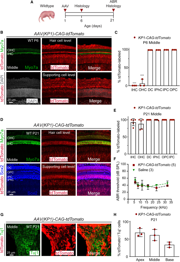
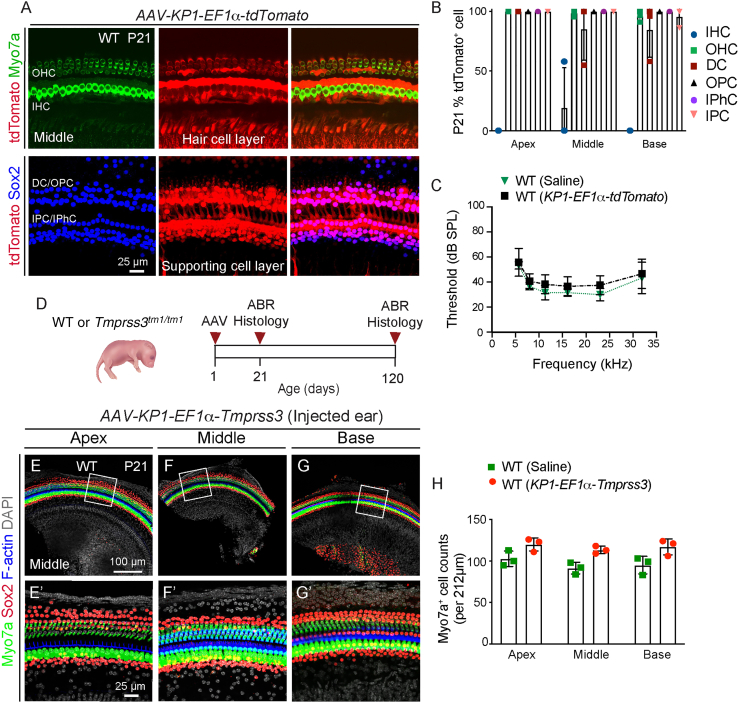
Adeno-associated virus (AAV)-mediated gene transfer has shown promise in rescuing mouse models of genetic hearing loss, but how viral capsid and promoter selection affects efficacy is poorly characterized. Here, we tested combinations of AAVs and promoters to deliver Tmprss3, mutations in which are associated with hearing loss in humans. Tmprss3tm1/tm1 mice display severe cochlear hair cell degeneration, loss of auditory brainstem responses, and delayed loss of spiral ganglion neurons. Under the ubiquitous CAG promoter and AAV-KP1 capsid, Tmprss3 overexpression caused striking cytotoxicity in vitro and in vivo and failed to rescue degeneration or dysfunction of the Tmprss3tm1/tm1 cochlea. Reducing the dosage or using AAV-DJ-CAG-Tmprss3 diminished cytotoxicity without rescue of the Tmprss3tm1/tm1 cochlea. Finally, the combination of AAV-KP1 capsid and the EF1α promoter prevented cytotoxicity and reduced hair cell degeneration, loss of spiral ganglion neurons, and improved hearing thresholds in Tmprss3tm1/tm1 mice. Together, our study illustrates toxicity of exogenous genes and factors governing rescue efficiency, and suggests that cochlear gene therapy likely requires precisely targeted transgene expression.

Keywords: Tmprss3; cochlea; gene therapy; hair cells; hearing loss; supporting cells.


Conflict of interest statement

K.P. and M.A.K. are inventors of filed patents held by Stanford University.

Figures

None
Graphical abstract
Figure 1
Figure 1
Tmprss3 deficiency causes cochlear hair cell degeneration and hearing loss (A) In situ hybridization (RNAScope) of P5 wild-type (WT) cochlea (middle turn and counterstained with hematoxylin) revealed Tmprss3 mRNA expression in hair cells and supporting cells, interdental cells, inner phalangeal cells, lesser epithelial ridge, outer sulcus, and Rosenthal’s canal. Schematic depicting hair cell and supporting cell subtypes. (B) The TMPRSS3 protein consists of 453 amino acids, with a transmembrane (TM) domain, a low-density lipoprotein receptor class A (LDRA), a scavenger receptor cysteine-rich domain (SRCR), and a C-terminal serine protease. The mutation was generated by targeted mutation through homologous recombination in exon 1. (C) At P21, Tmprss3tm1/+ littermates had ABR thresholds that were indistinguishable from WT littermates, whereas Tmprss3tm1/tm1 mice demonstrated no ABR responses across all frequencies tested. (D–F) Immunostaining of P12 cochleae showed no loss or disorganization of hair cells and supporting cells among WT, Tmprss3tm1/+, and Tmprss3tm1/tm1 littermates prior to the onset of hearing. (G–L) Substantial inner and outer hair cell loss and disorganized supporting cells were observed in the P21 and P120 Tmprss3tm1/tm1 mice. No cell loss in WT or Tmprss3tm1/+ cochleae. (M–N) Quantification in the middle cochlear turn showing significant loss of hair cells in P21 and P120 Tmprss3tm1/tm1 cochleae and medial supporting cell loss at P120. (O–Q) Cross sections of Rosenthal’s canal at P21 showing no spiral ganglia neuron degeneration. (R–T) At P120, there was a noticeable loss of spiral ganglion neurons in the Tmprss3tm1/tm1 cochleae. (U–V) Quantitative analysis showing a significant loss of TuJ1+ spiral ganglion neurons, but not Sox2+ glial cells, in P120 Tmprss3tm1/tm1 cochleae. Data shown as mean ± SD. ∗p < 0.05, ∗∗p < 0.01, ∗∗∗p < 0.001. Two-way ANOVA with Tukey’s multiple comparison. n = 3–6. IHC, inner hair cell; OHC, outer hair cell; DC, Deiters’ cell; IPhC, inner phalangeal cell; IPC, inner pillar cell; OPC, outer pillar cell; SGN, spiral ganglion neuron.
Figure 2
Figure 2
Tropism and efficacy of AAV-KP1-CAG-tdTomato in the cochlea (A) Schematic showing AAV injection at P1 in WT pups and examination at P6 and P21. (B) Robust tdTomato expression in supporting cells (bottom) but not hair cells (top) in the P6 cochlea (middle turn shown). (C) Quantification of labeled hair cells and supporting cells. (D) Robust tdTomato expression in both hair cells and supporting cells (top and bottom) at P21. (E) Quantitative analysis of tdTomato-labeled hair cells and supporting cell subtypes. (F) Both saline- and AAV-KP1-CAG-tdTomato-injected animals showed normal ABR thresholds at P21. (G) Only a subset of Tuj1+ spiral ganglion neurons were tdTomato labeled at P21. (H) Spiral ganglion neurons were partially transduced in all three cochlear turns, with the apex showing the highest rate. Data shown as mean ± SD. ∗∗∗p < 0.001. Two-way ANOVA with Tukey’s multiple comparison. n = 3–5.
Figure 3
Figure 3
Exogenous TMPRSS3 is cytotoxic in vitro and in vivo (A–C) Transduction of 293T/17 (human embryonic kidney) cells with AAV-KP1-CAG-Tmprss3 resulted in cell death in a dose-dependent manner, whereas none was observed in no-virus controls. (D) Proliferation assay demonstrating that increasing the MOI with AAV-KP1-CAG-Tmprss3 significantly decreased 293T/17 cell counts. Controls using huF9-expressing rAAV and no virus showed higher viability. (E) Schematic showing the timeline of injection of viral vectors into WT or mutant (Tmprss3tm1/tm1) cochleae and subsequent examination at P6 and P21. (F) At high titers (1:1, 2.0 × 108 vg), AAV-KP1-CAG-Tmprss3 caused degeneration of OHCs and swelling of IHCs. (G) A lower titer (1:2, 1.0 × 108 vg) did not cause hair cell degeneration, although IHCs still appeared swollen (inset). (H) No degeneration was observed at a 1:10 dilution (2.0 × 107 vg). (I–K) Hair cell degeneration was not prevented in P21 Tmprss3tm1/tm1 cochleae with any titers of AAV-KP1-CAG-Tmprss3 tested. (L) Quantification at P21 showing significant hair cell loss in all three turns of the cochlea at 1:1, but not other dilutions, of AAV-KP1-CAG-Tmprss3. (M) Hair cells degenerated in each turn of P21 Tmprss3tm1/tm1 cochleae injected with any titers of AAV-KP1-CAG-Tmprss3, resulting in significantly fewer hair cells than saline-injected, WT controls. (N) ABR thresholds were significantly higher in the ears of P21 WT animals injected with the full 1:1 titer AAV-KP1-CAG-Tmprss3 relative to saline-injected WT controls. (O and P) Some elevation of ABR thresholds was observed at a 1:2 dilution, and no changes were observed at a 1:10 dilution. Tmprss3tm1/tm1 mice displayed no ABR responses across all three titers. (Q) No cell loss was detected in the P21 WT cochlea after AAV-DJ-CAG-Tmprss3 had been injected at P1 (2.0 × 108 vg), although IHC appeared swollen. (R) Sensory hair cell loss at P21 was not prevented by AAV-DJ-CAG-Tmprss3 in Tmprss3tm1/tm1 mice. (S) Hair cell counts in saline- and AAV-DJ-CAG-Tmprss3-injected WT cochleae and AAV-DJ-CAG-Tmprss3-injected Tmprss3tm1/tm1 cochleae. (T) WT ears injected with AAV-DJ-CAG-Tmprss3 showed elevated ABR thresholds at 8 and 16 kHz. Tmprss3tm1/tm1 ears treated with AAV-DJ-CAG-Tmprss3 had no ABR responses. Data shown as mean ± SD. ∗p < 0.05, ∗∗p < 0.01, ∗∗∗p < 0.001. Two-way ANOVA with Tukey’s multiple comparison. n = 3.
Figure 4
Figure 4
AAV-KP1-EF1α-Tmprss3 is not cytotoxic in the cochlea in vivo (A) After injection of AAV-KP1-EF1α-tdTomato (1.0 × 109 vg/mL) at P1, there was robust tdTomato expression in OHCs (top) and supporting cell subtypes (lower) at P21 (middle turn shown). (B) Most OHCs and supporting cell subtypes and almost no IHCs were tdTomato labeled. (C) Relative to saline-injected ears, there were no detectable shifts in ABR thresholds in the AAV-KP1-EF1α-tdTomato-injected ears at P21. (D) Schematic showing the timeline of injection of AAV-KP1-EF1α-Tmprss3 into WT or mutant (Tmprss3tm1/tm1) cochleae and subsequent examination at P21 and P120. (E–G) AAV-KP1-EF1α-Tmprss3 (6.5 × 108 vg) injected into P1 WT mice did not cause any loss of sensory cells or supporting cells across all three turns at P21. (E′–G′) High-magnification images from (E–G). (H) Quantification of hair cells in saline- and AAV-KP1-EF1α-Tmprss3-injected WT cochleae. Data shown as mean ± SD. Two-way ANOVA with Tukey’s multiple comparison. n = 3.
Figure 5
Figure 5
AAV-KP1-EF1α-Tmprss3 partially prevents degeneration and auditory dysfunction in Tmprss3tm1/tm1 mice (A–C) After AAV-KP1-EF1α-Tmprss3 injection (6.5 × 108 vg) in P1 Tmprss3tm1/tm1 mice, most IHCs and OHCs were present in the middle and basal turns, while most IHCs and some OHCs remained in the apical turn at P21. (A′–C′) High-magnification images from (A)–(C). (D) Myo7a+ cell counts in each turn of treated Tmprss3tm1/tm1 cochlea were significantly higher than untreated Tmprss3tm1/tm1 cochlea and were similar to WT controls. (E–G) Hair cell survival persisted in each turn of P120-treated Tmprss3tm1/tm1 cochlea. (H) Each turn of P120-treated Tmprss3tm1/tm1 cochlea displayed significantly more hair cells than untreated Tmprss3tm1/tm1 cochlea and similar to WT controls. (I–K) Many SGNs were preserved in each turn of P120-treated Tmprss3tm1/tm1 cochlea, particularly the middle and basal turns. (L) Treated Tmprss3tm1/tm1 cochlea had higher SGN counts than untreated Tmprss3tm1/tm1 cochlea. (M) Raw ABR waveforms of P21 WT, untreated, and treated Tmprss3tm1/tm1 mice at 16kHz. (N and O). All treated Tmprss3tm1/tm1 ears demonstrated detectable ABR responses at P21 and P120, whereas untreated ears showed no responses. Data shown as mean ± SD. ∗p < 0.05, ∗∗p < 0.01, ∗∗∗p < 0.001, ∗∗∗∗p < 0.0001. Two-way ANOVA with Tukey’s multiple comparison. n = 3–5.

References

    1. Morton C.C., Nance W.E. Newborn hearing screening--a silent revolution. N. Engl. J. Med. 2006;354:2151–2164. doi: 10.1056/NEJMra050700. - DOI - PubMed
    1. Duman D., Tekin M. Autosomal recessive nonsyndromic deafness genes: a review. Front. Biosci. 2012;17:2213–2236. - PMC - PubMed
    1. Scott H.S., Kudoh J., Wattenhofer M., Shibuya K., Berry A., Chrast R., Guipponi M., Wang J., Kawasaki K., Asakawa S., et al. Insertion of beta-satellite repeats identifies a transmembrane protease causing both congenital and childhood onset autosomal recessive deafness. Nat. Genet. 2001;27:59–63. - PubMed
    1. Wattenhofer M., Sahin-Calapoglu N., Andreasen D., Kalay E., Caylan R., Braillard B., Fowler-Jaeger N., Reymond A., Rossier B.C., Karaguzel A., Antonarakis S.E. A novel TMPRSS3 missense mutation in a DFNB8/10 family prevents proteolytic activation of the protein. Hum. Genet. 2005;117:528–535. doi: 10.1007/s00439-005-1332-x. - DOI - PubMed
    1. Seligman K.L., Shearer A.E., Frees K., Nishimura C., Kolbe D., Dunn C., Hansen M.R., Gantz B.J., Smith R.J.H. Genetic Causes of Hearing Loss in a Large Cohort of Cochlear Implant Recipients. Otolaryngol. Head Neck Surg. 2022;166:734–737. doi: 10.1177/01945998211021308. - DOI - PMC - PubMed

LinkOut - more resources