Review
. 2023 Aug 17:10:1213320.
doi: 10.3389/fmed.2023.1213320. eCollection 2023.

Mast cell-mediated immune regulation in health and disease

Affiliations
Review

Mast cell-mediated immune regulation in health and disease

Kottarappat N Dileepan et al. Front Med (Lausanne). .

Abstract

Mast cells are important components of the immune system, and they perform pro-inflammatory as well as anti-inflammatory roles in the complex process of immune regulation in health and disease. Because of their strategic perivascular localization, sensitivity and adaptability to the microenvironment, and ability to release a variety of preformed and newly synthesized effector molecules, mast cells perform unique functions in almost all organs. Additionally, Mast cells express a wide range of surface and cytoplasmic receptors which enable them to respond to a variety of cytokines, chemicals, and pathogens. The mast cell's role as a cellular interface between external and internal environments as well as between vasculature and tissues is critical for protection and repair. Mast cell interactions with different immune and nonimmune cells through secreted inflammatory mediators may also turn in favor of disease promoting agents. First and forefront, mast cells are well recognized for their multifaceted functions in allergic diseases. Reciprocal communication between mast cells and endothelial cells in the presence of bacterial toxins in chronic/sub-clinical infections induce persistent vascular inflammation. We have shown that mast cell proteases and histamine induce endothelial inflammatory responses that are synergistically amplified by bacterial toxins. Mast cells have been shown to exacerbate vascular changes in normal states as well as in chronic or subclinical infections, particularly among cigarette smokers. Furthermore, a potential role of mast cells in SARS-CoV-2-induced dysfunction of the capillary-alveolar interface adds to the growing understanding of mast cells in viral infections. The interaction between mast cells and microglial cells in the brain further highlights their significance in neuroinflammation. This review highlights the significant role of mast cells as the interface that acts as sensor and early responder through interactions with cells in systemic organs and the nervous system.

Keywords: SARS-CoV-2 disease; allergic disease; atherosclerosis; bacterial infection; cardiovascular disease; cigarette smoking; neuroinflammation; skin and wound-healing.


Conflict of interest statement

The authors declare that the research was conducted in the absence of any commercial or financial relationships that could be construed as a potential conflict of interest.

Figures
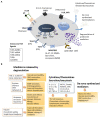
Figure 1
Figure 1
Mast cell receptors and products. Panel (A) depicts some of the major receptors expressed on mast cells. Respective ligands of these receptors are also shown. The receptors shown here include the high affinity FceRI for IgE; MRGPRX2: Mas-related G-protein coupled receptor-X2 that binds quinolones and compound 48/80 among other ligands; TLRs (Toll-like receptors) at the plasma membrane (TLR2 and TLR4) and at endosomes (TLR3 and TLR7-9) that bind to various exogenous and endogenous molecules in a pattern recognizing manner; NPR for neuropeptide ligands ANP, BNP or CNP; Protease Activated Receptors PARs; Histamine Receptors HRs for histamine; CXC-Chemokine Receptor CXCR. Other important receptors expressed on mast cells (not shown here) include receptors for Kit (stem cell factor receptor), Complement (C3a and C5a); alarmins IL-33 (ST2, IL-1 family receptor) and Epithelial cell derived Thymic stromal lymphopoietin (TSLP); the platelet-activating factor (PAF); vascular endothelial growth factor (VEGF); nerve growth factor (NGF); fibroblast growth factor (FGF); Interleukins (IL); Transforming growth factor beta (TGF-b); Thymus and activation-regulated chemokine (TARC); Prostaglandins (PGs); Cysteinyl leukotrienes (CysLT). Mast cell regulator/inhibitory receptors containing immunoregulatory tyrosine inhibition motifs (ITIMs) including IgE receptor Fc-gamma-RIIb, CD300a, CD200 R1, platelet-endothelial cell adhesion molecule 1 (PECAM-1), paired immunoglobulin-like receptor B (PIR-B), the c-lectin mast cell function-associated antigen (MAFA), sialic acid-binding immunoglobulin-like lectins (Siglecs), and leukocyte immunoglobulin-like receptor 4, subfamily B, member 4 (LILRB4). Panel (B) mast cells release many effector molecules through different mechanisms. Degranulation is a robust mechanism for mast cells to release pre-formed molecules of several chemical classes. However, release of mediators through degranulation is a complex process that may vary with regard to the composition of granules or the duration of release resulting in a fine control of mast cell response to their surroundings. Release of molecules including cytokines and chemokines may involve the classical secretory pathway or other mechanism for exocytosis. Molecules such as prostaglandins and leukotrienes are synthesized de novo in response to stimuli. Mediators released by mast cells enable mast cells to respond to many physiological and pathological conditions. bFGF, Basic Fibroblast Growth Factor; CCL2,3, and 4, Chemokine (C-C motif) Ligand 2, 3 and 4; CPA3, Carboxypeptidase-3; CXCL2,3 and 4-Chemokine (C-X-C motif) Ligand 2, 3 and 4; cysLTs, Cystinyl Leukotrienes; GM-CSF, Granulocyte Macrophage Colony-Stimulating Factor; IL-1, IL-3, IL-4, IL-6, IL-18, Interleukins 1, 3, 4, 6, 18; NGF, Nerve Growth Factor; MCP-1, Monocyte chemoattractant protein-1 (CCL2); PAF, Platelet Activating Factor; PGs, Prostaglandins; RANTES, Regulated upon Activation, Normal T cell Expressed and Secreted aka (CCL5); SCF, Stem Cell Factor; SMC, Smooth Muscle cells; TARC, Thymus and Activation Regulated Chemokine; TGF-β, Transforming Growth factor-β; TNF, Tumor Necrosis Factor; VEGF, Vascular Endothelial Growth Factor/vascular permeability factor.
Figure 2
Figure 2
Schematic diagram showing the effect of mast cell-derived compounds on specific cell types. Mast cells have stimulatory or inhibitory actions on many types of cells. A few of those cell types are shown in this diagram. For the simplicity, the scheme shows only most studied effector molecules.
Figure 3
Figure 3
Schematic representation of the potential role of mast cells in SARS-CoV-12- induced tissue/organ damage. Spike protein of SARS-CoV-2 binds to ACE2 receptor on mast cells to cause degranulation. The hyperactive mast cells-derived granules contain various bioactive molecules that affect organ systems as mentioned. Notably, mast cell activation is now considered to be a key mechanism involved in various organ-related symptoms indicating long COVID.

References

    1. Turvey SE, Broide DH. Innate immunity. J Allergy Clin Immunol. (2010) 125:S24–32. doi: 10.1016/j.jaci.2009.07.016, PMID: - DOI - PMC - PubMed
    1. Krystel-Whittemore M, Dileepan KN, Wood JG. Mast cell: a multi-functional master cell. Front Immunol. (2015) 6:620. doi: 10.3389/fimmu.2015.00620, PMID: - DOI - PMC - PubMed
    1. Ehrlich P. Beitraege Zur Kenntniss Der Anilinfaerbungen Und Ihrer Verwendung in Der Mikroskopischen Technik. Arch mikr anat u entwcklngsmech. (1877) 13:263–77. doi: 10.1007/BF02933937 - DOI
    1. Saito H, Matsumoto K, Okumura S, Kashiwakura J-i, Oboki K, Yokoi H, et al. . Gene expression profiling of human mast cell subtypes: an in silico study. Allergol Int. (2006) 55:173–9. doi: 10.2332/allergolint.55.173, PMID: - DOI - PubMed
    1. Quackenbush EJ, Wershil BK, Aguirre V, Gutierrez-Ramos JC. Eotaxin modulates Myelopoiesis and mast cell development from embryonic hematopoietic progenitors. Blood. (1998) 92:1887–97. doi: 10.1182/blood.V92.6.1887, PMID: - DOI - PubMed