. 2023 Oct;13(10):323.
doi: 10.1007/s13205-023-03749-y. Epub 2023 Aug 30.

Understanding the role of conserved proline and serine residues in the SARS-CoV-2 spike cleavage sites in the virus entry, fusion, and infectivity

Affiliations

Understanding the role of conserved proline and serine residues in the SARS-CoV-2 spike cleavage sites in the virus entry, fusion, and infectivity

Ritika Khatri et al. 3 Biotech. 2023 Oct.

Abstract

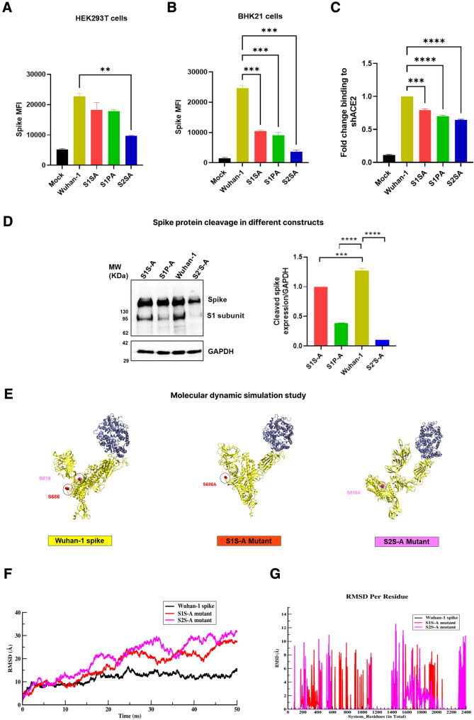
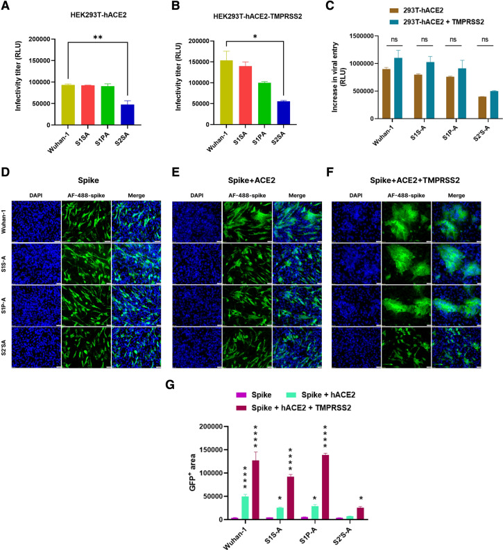
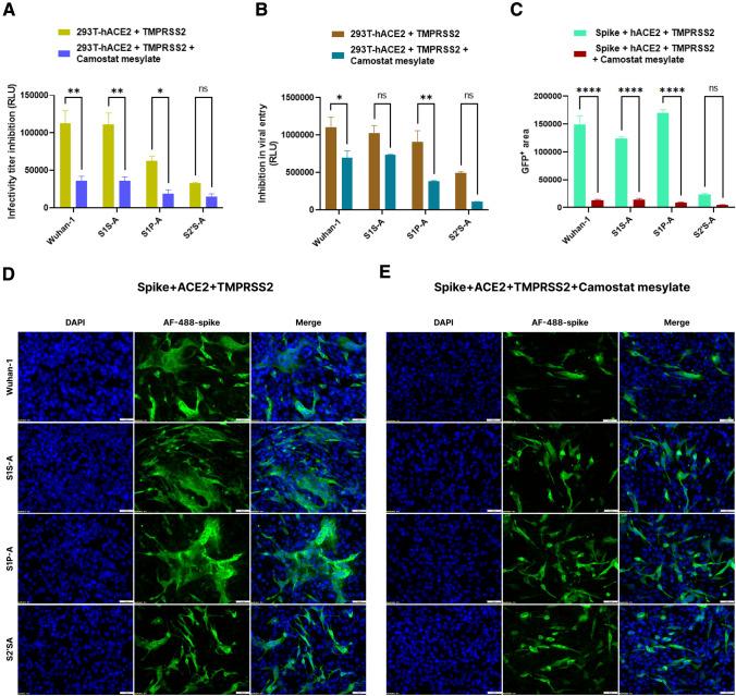
The spike (S) glycoprotein of the SARS-CoV-2 virus binds to the host cell receptor and promotes the virus's entry into the target host cell. This interaction is primed by host cell proteases like furin and TMPRSS2, which act at the S1/S2 and S2´ cleavage sites, respectively. Both cleavage sites have serine or proline residues flanking either the single or polybasic region and were found to be conserved in coronaviruses. Unravelling the effects of these conserved residues on the virus entry and infectivity might facilitate the development of novel therapeutics. Here, we have investigated the role of the conserved serine and proline residues in the SARS-CoV-2 spike mediated entry, fusogenicity, and viral infectivity by using the HIV-1/spike-based pseudovirus system. A conserved serine residue mutation to alanine (S2´S-A) at the S2´ cleavage site resulted in the complete loss of spike cleavage. Exogenous treatment with trypsin or overexpression of TMPRSS2 protease could not rescue the loss of spike cleavage and biological activity. The S2´S-A mutant showed no significant responses against E-64d, TMPRSS2 or other relevant inhibitors. Taken together, serine at the S2´ site in the spike protein was indispensable for spike protein cleavage and virus infectivity. Thus, novel interventions targeting the conserved serine at the S2´ cleavage site should be explored to reduce severe disease caused by SARS-CoV-2-and novel emerging variants.

Supplementary information: The online version contains supplementary material available at 10.1007/s13205-023-03749-y.

Keywords: Cell entry and viral infectivity; Conserved serine; SARS-CoV-2; Spike cleavage site.


Conflict of interest statement

Conflict of interestOn behalf of all authors, the corresponding author states that there is no conflict of interest.

Figures

Fig. 1
Fig. 1
Schematic presentation of conserved residues and cleavage site mutations in spike. A Conserved residues are shown in spike protein of different coronaviruses around the S1/S2 furin and S2´ cleavage sites. Cleavage site amino acids (arginine, R) are in red bold font and the conserved residues (serine, S and proline, P) are in blue bold font. RBD, Receptor binding domain; FP, fusion peptide; N, N-terminal region and C, C-terminal region. Coronaviruses: SARS-CoV-2, severe acute respiratory syndrome coronavirus-2; SARS-CoV, severe acute respiratory syndrome coronavirus-1; MERS CoV, Middle East respiratory syndrome coronavirus; HCoV, human coronavirus; and IBV, infectious bronchitis virus. B Cleavage site amino acid residues in SARS-CoV-2 spike. Conserved serine and proline residues are shown in bold black font. Mutations in the conserved serine and proline residues to alanine: S1S-A, S1/S2 site serine to alanine mutation; S1P-A, S1/S2 site proline to alanine mutation and S2´S-A, S2´ cleavage site serine to alanine mutant) are shown in blue bold font. SP signal peptide, NTD N-terminal domain, RBD receptor binding domain, FP fusion peptide, HR1 heptad repeat 1, HR2 heptad repeat 2, TM transmembrane domain and CP cytoplasmic tail
Fig. 2
Fig. 2
Spike expression, ACE2 binding and cleavage of cleavage site mutants. A, B Cell surface expression of spike protein variants in HEK293T and BHK-21 cell lines as measured by flow cytometry. BHK-21 and HEK293T cells were transfected with spike cleavage site mutants for 24 h. Post incubation, spike expressing cells were stained with polyclonal mice anti-spike sera and later, PE-conjugated anti-mouse goat secondary antibody. Samples were analyzed with BD FACS Diva flow cytometer. Statistical significance was determined using one-way Anova in GraphPad prism keeping Wuhan-1 as the control. The experiment was repeated three times and the error bars show mean values with SEM. C Soluble-hACE2 protein binding with spike cleavage site mutants. Spike protein was expressed on HEK293T cell surface and after 24 h of transfection, cells were incubated with 10ug of soluble-hACE2 for an hour, stained with anti-hACE2 mouse polyclonal sera and later with PE-conjugated anti-mouse goat secondary antibody. Samples were acquired with BD FACS Diva flow cytometer. Statistical significance was determined using one-way Anova in GraphPad prism keeping Wuhan-1 as the control. The experiment was repeated three times and the error bars show mean values with SEM. D Western blot analysis of spike expression in HEK293T cells at 24 h post transfection. Cell lysates were prepared using RIPA lysis buffer and run on a 10% SDS-PAGE. After transferring the gel to a PVDF membrane, it was blocked with 5% milk and stained with anti-RBD mouse polyclonal sera (1:1000 dilution) and later with HRP-conjugated goat anti-mouse antibody (1:2000). GAPDH was used as a loading control. Experiment was repeated three times and the error bar show mean values with SEM. For the statistical significance, one-way ANOVA test was performed with multiple comparisons where, Wuhan-1 was used as a control to compare with cleavage site mutants. (p < 0.05), where (p < 0.05), *P < 0.05, **P < 0.01, ***P < 0.001, ****P < 0.0001 were considered significant and p > 0.05 was considered nonsignificant (ns). EG Dynamical measurement of structures over time. E Conformational changes in the protein-complex of spike protein with hACE2. Figure shows hACE2 protein complex with Wuhan-1, S1S-A, and S2´SA, respectively. F RMSD analysis of Wuhan-1/mutant spike with hACE2 protein complex system over the course of MD (molecular dynamics) trajectory simulation. G Changes in RMSD at residual level for S1S-A and S2´S-A mutants in comparison with Wuhan-1 spike
Fig. 3
Fig. 3
Effect of ACE2 and TMPRSS2 on the infectivity, viral entry and fusogenicity of cleavage site mutants. A, B Infectivity titer of pseudovirus variants in HEK293T-hACE2 and HEK293T-hACE2-TMPRSS2 cells, respectively. Spike cleavage site mutants based pseudoviruses were prepared in HEK293T cells. Then, HEK293T-hACE2 and HEK293T-hACE2-TMPRSS2 cells were infected with pseudoviruses for 48 h. Cells were lysed and luciferase titer was measured in a luminometer. Statistical significance was determined using one-way Anova keeping Wuhan-1 as the control. The experiment was repeated three times and the error bars show mean values with SEM. C Quantitative fusion assay of spike variants with HEK293T-hACE2 or HEK293T-hACE2-TMPRSS2 as measured by RLU. The experiment was repeated three times in duplicates. Statistical significance was determined using two-way ANOVA, with multiple comparisons using hACE2 expression as control. The error bars show mean values with SEM. DF Immunofluorescence assay for spike (D), spike-hACE2 (E) and spike-hACE2-TMPRSS2 (F) mediated cell–cell fusion in BHK-21 cells at 24 h post transfection, respectively. After transfecting the cells with respective plasmids, cells were fixed post 24-h and stained with anti-spike mouse polyclonal sera (1:500) and AlexaFluor-488-labeled anti-mouse antibody (green) (1:1000). Experiment was repeated three times. The images were taken with an Olympus fluorescence microscope. The nuclei were stained with 4′,6-diamidino-2-phenylindole (DAPI, blue); scale bar: 50 μm and magnification 20x. G Immunofluorescence images were quantified by selecting five random GFP + areas in Image J software. Graph was prepared and analyzed in GraphPad prism 9. For the statistical significance two-way ANOVA test was performed with multiple comparisons where, spike expression was used as a control to compare with spike + hACE2 or spike + hACE2 + TMPRSS2 expression. The error bars show mean values with SEM. (p < 0.05), *P < 0.05, **P < 0.01, ***P < 0.001, ****P < 0.0001 were considered significant and p > 0.05 was considered nonsignificant (ns)
Fig. 4
Fig. 4
Effect of E-64d and NH4Cl on the infectivity, viral entry and fusogenicity of cleavage site mutants. A Inhibition in infectivity titer of pseudovirus variants in HEK293T-hACE2 in the presence of E-64d. After harvesting, the pseudoviruses were incubated with 20 µM of E-64d for 2 h at 37 ºC. Later, HEK293T-hACE2 cells were added to pseudovirus and drug mixture. Post 48 h of cell addition, luciferase readings were taken as RLU. Statistical significance was determined using two-way ANOVA, with multiple comparisons using spike + hACE2 (untreated) as control. The error bars show mean values with SEM. The experiment was repeated three times. B Inhibition in viral entry of pseudovirus variants in HEK293T-hACE2 in the presence of E-64d. 20 µM of cysteine protease inhibitor, E-64d, was added to the entry assay cells for 2 h at 37ºC. Later, HEK293T-hACE2 cells were added to the spike expressing cells and drug compound. Post 48 h of cell addition, luciferase readings were taken as RLU. Statistical significance was determined using two-way ANOVA, with multiple comparisons using spike + hACE2 (untreated) as control. The error bars show mean values with SEM. The experiment was repeated three times. CE Immunofluorescence assay for fusion inhibition in spike variants of SARS-CoV-2 in BHK-21 cells in the presence of E-64d at 24 h post transfection. The experiment was repeated three times. Statistical significance was determined using two-way ANOVA, with multiple comparisons using hACE2 expression without E-64d (D) as control. The error bars show mean values with SEM. F. Immunofluorescence assay for fusion inhibition in spike variants of SARS-CoV-2 in BHK-21 cells in the presence of NH4Cl (20 mM) at 24 h post transfection. After transfecting the cells with respective plasmids, cells were treated with compound post 2 h of transfection. Later, cells were fixed post 24-h and stained with anti-spike mouse polyclonal sera (1:500) and AlexaFluor-488-labeled anti-mouse antibody (green) (1:1000). Experiment was repeated three times. The images were taken with an Olympus fluorescence microscope. The nuclei were stained with 4′,6-diamidino-2-phenylindole (DAPI, blue); scale bar: 50 μm and magnification 20x. G. Inhibition in infectivity titer of pseudovirus variants in HEK293T-hACE2 in the presence of NH4Cl at 48 h of infection. H Inhibition in viral entry of pseudovirus variants in HEK293T-hACE2 in the presence of NH4Cl. Experiment was repeated three times. For the statistical significance, two-way ANOVA test was performed with multiple comparisons where untreated samples were compared with NH4Cl treated samples. The error bars show mean values with SEM. I Immunofluorescence images for NH4Cl treatment were quantified by selecting five random GFP + areas in Image J software. Graph was prepared and analyzed in GraphPad prism 9. For the statistical significance two-way ANOVA test was performed with multiple comparisons. p < 0.05, *P < 0.05, **P < 0.01, ***P < 0.001, and ****P < 0.0001 were considered significant and p > 0.05 was considered nonsignificant (ns)
Fig. 5
Fig. 5
Effect of camostat mesylate on the infectivity, viral entry and fusogenicity of cleavage site mutants. A Inhibition in infectivity titer of pseudovirus variants in HEK293T-hACE2-TMPRSS2 cells in the presence of camostat mesylate (100 µM) at 48 h of infection. Experiment was repeated three times. The error bars show mean values with SEM. For the statistical significance, two-way ANOVA test was performed with multiple comparisons where untreated samples were compared with camostat mesylate treated samples. B Inhibition in cell entry of pseudovirus variants in HEK293T-hACE2-TMPRSS2 cells in the presence of camostat mesylate. Camostat mesylate (100 µM) was added to the entry assay cells for 2 h at 37 ºC. Later, HEK293T-hACE2 cells were added to the spike expressing cells and drug compound. Post 48 h of cell addition, luciferase readings were taken as RLU. The error bars show mean values with SEM. Experiment was repeated three times. For the statistical significance, two-way ANOVA test was performed with multiple comparisons where untreated samples were compared with camostat mesylate treated samples. CE. Immunofluorescence assay for fusion inhibition mediated by spike, hACE2 and TMPRSS2 in BHK-21 cells in the presence of camostat mesylate at 24 h post transfection. After transfecting the cells with respective plasmids, cells were treated with compound post 2 h of transfection. Later, cells were fixed post 24-h and stained with anti-spike mouse polyclonal sera (1:500) and AlexaFluor-488-labeled anti-mouse antibody (green) (1:1000). Experiment was repeated three times. The images were taken with an Olympus fluorescence microscope. The nuclei were stained with 4′,6-diamidino-2-phenylindole (DAPI, blue); scale bar: 50 μm and magnification 20x. The error bars show mean values with SEM. For the statistical significance, two-way ANOVA test was performed with multiple comparisons where untreated samples were compared with camostat mesylate treated samples. p < 0.05, *P < 0.05, **P < 0.01, ***P < 0.001, and ****P < 0.0001 were considered significant and p > 0.05 was considered nonsignificant (ns)
Fig. 6
Fig. 6
Effect of batimastat and cytochalasin D on the infectivity, viral entry and fusogenicity of cleavage site mutants. A, B Immunofluorescence assay for fusion inhibition in the spike variants of SARS-CoV-2 in BHK-21 cells in the presence of batimastat (20 µM) at 24 h post transfection. Batimastat was added to the spike-hACE2 and spike-hACE2-TMPRSS2 expressing cells. After transfecting the cells with respective plasmids, cells were treated with compound post 2 h of transfection. Later, cells were fixed post 24-h and stained with anti-spike mouse polyclonal sera (1:500) and AlexaFluor 488-labeled anti-mouse antibody (green) (1:1000). Experiment was repeated three times. The images were taken with an Olympus fluorescence microscope. The nuclei were stained with 4′,6-diamidino-2-phenylindole (DAPI, blue); scale bar: 50 μm and magnification 20x. C Inhibition in infectivity titer of pseudovirus variants in HEK293T-hACE2 and HEK293T-hACE2-TMPRSS2 cells in the presence of batimastat at 48 h post infection. The error bars show mean values with SEM. Experiment was repeated three times. For the statistical significance, two-way ANOVA test was performed with multiple comparisons. D, E Immunofluorescence images for batimastat treatment were quantified by selecting five random GFP + areas in Image J software. Graph was prepared and analyzed in GraphPad prism 9. The error bars show mean values with SEM. For the statistical significance, two-way ANOVA test was performed with multiple comparisons where untreated samples (Fig. 4D) were compared with batimastat treated samples in the presence or absence of TMPRSS2. F, G Immunofluorescence assay for fusion inhibition in spike variants of SARS-CoV-2 in BHK-21 cells in the presence of cytochalasin D (1 µM) at 24 h post transfection. Cytochalasin D was added to the spike-hACE2 and spike-hACE2-TMPRSS2 expressing cells. After transfecting the cells with respective plasmids, cells were treated with compound post 2 h of transfection. Later, cells were fixed post 24-h and stained with anti-spike mouse polyclonal sera (1:500) and AlexaFluor-488-labeled anti-mouse antibody (green) (1:1000). Experiment was repeated three times. The images were taken with an Olympus fluorescence microscope. The nuclei were stained with 4′,6-diamidino-2-phenylindole (DAPI, blue); scale bar: 50 μm and magnification 20x. H, I Inhibition in infectivity titer of pseudovirus variants in HEK293T-hACE2 and HEK293T-hACE2-TMPRSS2 cells in the presence of cytochalasin D at 48 h post infection. The error bars show mean values with SEM. Experiment was repeated three times. For the statistical significance, two-way ANOVA test was performed with multiple comparisons. I Immunofluorescence images for cytochalasin D treatment were quantified by selecting five random GFP + areas in Image J software. Graph was prepared and analyzed in GraphPad prism 9. The error bars show mean values with SEM. For the statistical significance, two-way ANOVA test was performed with multiple comparisons where untreated samples (Fig. 4D) were compared with cytochalasin D treated samples in the presence or absence of TMPRSS2. J. Inhibition in infectivity titer of pseudovirus variants in HEK293T-hACE2 cells in the presence of cytochalasin D at 48 h post infection. Experiment was repeated three times. For the statistical significance, two-way ANOVA test was performed with multiple comparisons where untreated samples were compared with cytochalasin D treated samples. p < 0.05, *P < 0.05, **P < 0.01, ***P < 0.001, and ****P < 0.0001 were considered significant and p > 0.05 was considered nonsignificant (ns)

Similar articles

Cited by

References

    1. Adedeji AO, Severson W, Jonsson C, et al. Novel inhibitors of severe acute respiratory syndrome coronavirus entry that act by three distinct mechanisms. J Virol. 2013;87:8017–8028. doi: 10.1128/JVI.00998-13. - DOI - PMC - PubMed
    1. Alaofi AL, Shahid M. Mutations of SARS-CoV-2 RBD may alter its molecular structure to improve its infection efficiency. Biomolecules. 2021 doi: 10.3390/biom11091273. - DOI - PMC - PubMed
    1. Amitai A. Viral surface geometry shapes influenza and coronavirus spike evolution through antibody pressure. PLoS Comput Biol. 2021;17:e1009664. doi: 10.1371/journal.pcbi.1009664. - DOI - PMC - PubMed
    1. Arora P, Sidarovich A, Krüger N, et al. B.1.617.2 enters and fuses lung cells with increased efficiency and evades antibodies induced by infection and vaccination. Cell Rep. 2021;37:109825. doi: 10.1016/j.celrep.2021.109825. - DOI - PMC - PubMed
    1. Belouzard S, Chu VC, Whittaker GR. Activation of the SARS coronavirus spike protein via sequential proteolytic cleavage at two distinct sites. Proc Natl Acad Sci U S A. 2009;106:5871–5876. doi: 10.1073/pnas.0809524106. - DOI - PMC - PubMed

LinkOut - more resources