. 2023 Aug 31;3(8):1716-1730.
doi: 10.1158/2767-9764.CRC-23-0208. eCollection 2023 Aug.

Demethylation of EHMT1/GLP Protein Reprograms Its Transcriptional Activity and Promotes Prostate Cancer Progression

Affiliations

Demethylation of EHMT1/GLP Protein Reprograms Its Transcriptional Activity and Promotes Prostate Cancer Progression

Anna Besschetnova et al. Cancer Res Commun. .

Abstract

Epigenetic reprogramming, mediated by genomic alterations and dysregulation of histone reader and writer proteins, plays a critical role in driving prostate cancer progression and treatment resistance. However, the specific function and regulation of EHMT1 (also known as GLP) and EHMT2 (also known as G9A), well-known histone 3 lysine 9 methyltransferases, in prostate cancer progression remain poorly understood. Through comprehensive investigations, we discovered that both EHMT1 and EHMT2 proteins have the ability to activate oncogenic transcription programs in prostate cancer cells. Silencing EHMT1/2 or targeting their enzymatic activity with small-molecule inhibitors can markedly decrease prostate cancer cell proliferation and metastasis in vitro and in vivo. In-depth analysis of posttranslational modifications of EHMT1 protein revealed the presence of methylation at lysine 450 and 451 residues in multiple prostate cancer models. Notably, we found that lysine 450 can be demethylated by LSD1. Strikingly, concurrent demethylation of both lysine residues resulted in a rapid and profound expansion of EHMT1's chromatin binding capacity, enabling EHMT1 to reprogram the transcription networks in prostate cancer cells and activate oncogenic signaling pathways. Overall, our studies provide valuable molecular insights into the activity and function of EHMT proteins during prostate cancer progression. Moreover, we propose that the dual-lysine demethylation of EHMT1 acts as a critical molecular switch, triggering the induction of oncogenic transcriptional reprogramming in prostate cancer cells. These findings highlight the potential of targeting EHMT1/2 and their demethylation processes as promising therapeutic strategies for combating prostate cancer progression and overcoming treatment resistance.

Significance: In this study, we demonstrate that EHMT1 and EHMT2 proteins drive prostate cancer development by transcriptionally activating multiple oncogenic pathways. Mechanistically, the chromatin binding of EHMT1 is significantly expanded through demethylation of both lysine 450 and 451 residues, which can serve as a critical molecular switch to induce oncogenic transcriptional reprogramming in prostate cancer cells.


Figures

FIGURE 1
FIGURE 1
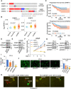
EHMT1 and EHMT2 promote prostate cancer (PCa) development. A,EHMT1 and EHMT2 gene alterations in two prostate cancer cohorts (TCGA primary prostate cancer and SU2C mCRPC). B, Box plots depicting the expression levels of EHMT1 or EHMT2 in normal and primary prostate cancer samples (TCGA) and mCRPC samples (SU2C). C, Kaplan–Meier curve illustrating the overall survival in prostate cancer tumors with higher EHMT1 or EHMT2 expression (red, the top 25%) compared with those with lower expression (blue, the bottom 75%). Immunoblotting analysis of indicated proteins in LNCaP cells transfected with siRNA pools against EHMT1 (D) or EHMT2 (E) for 2 days. F, Proliferation assay conducted on LNCaP cells transfected with siEHMT1, siEHMT2, or the combination for 0–4 days. Immunoblotting analysis of indicated proteins in PC-3 cells transfected with siEHMT1 (G) or siEHMT2 (H) for 2 days. Proliferation assay (I) and transwell migration assay (J) performed on PC-3 cells transfected with siEHMT1, siEHMT2, or the combination for 4 days (proliferation) and 3 days (migration). K, PC-3 cells (stably expressing GFP) transfected with siNTC versus siEHMT1 (for 3 days) were injected into zebrafish embryos (N = 10), and tumor cell intravasation was observed using a fluorescence microscope. The number of embryos showing invasion signals was counted. TCGA, The Cancer Genome Atlas.
FIGURE 2
FIGURE 2
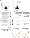
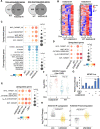
EHMT1 and EHMT2 transcriptionally activate oncogenic programs. A and B, RNA-seq analyses were conducted in LNCaP cells transfected with siEHMT1, siEHMT2, or siNTC for 2 days. Volcano plots show genes regulated by EHMT1 (A) or EHMT2 (B). C,GSEA of EHMT1 or EHMT2-upregulated genes (siEHMT1 or siEHMT2-downregulated) for hallmark gene set (cutoff: Padj < 0.05). D, GSEA of EHMT1-regulated genes to assess the enrichment of LSD1 function, based on previously determined LSD1 target gene sets (32) and using RNA-seq from GSE52201. E, GSEA of EHMT2-regulated genes to evaluate the enrichment of LSD1 function. F, Box plots presenting the expression levels of EHMT1-activated or EHMT2-activated genes (cutoff: 2-fold, P < 0.05) in normal and primary prostate cancer samples (TCGA) as well as mCRPC samples (SU2C). TCGA, The Cancer Genome Atlas.
FIGURE 3
FIGURE 3
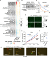
A specific EHMT1/2 dual inhibitor suppresses prostate cancer growth and metastasis in vitro and in vivo.A, GSEA conducted on differentially regulated genes by UNC0642 (2 μmol/L, 2 days), siEHMT1, or siEHMT2 (based on RNA-seq analyses). B, Immunoblotting analysis of indicated proteins in LNCaP or PC-3 cells treated with UNC0642 (0, 2, 10 μmol/L, 2 days). C, Proliferation assay performed on LNCaP or PC-3 cells treated with UNC0642 (0–20 μmol/L, 3 days). D, Transwell migration assay conducted on LNCaP or PC-3 cells treated with UNC0642 (1 μmol/L, 2 days). E, PC-3 cells were subcutaneously injected into male SCID mice. Once the tumor was established, mice (N = 6) were treated with UNC0642 (10 mg/kg) via oral gavage, and tumor volume was measured by caliper. F, Normalized H3K9me2 expression was calculated (using ImageJ for quantification) based on immunoblotting of H3K9me2 and GAPDH in tumor samples. G, PC-3 cells (stably expressing GFP) pretreated with UNC0642 (2 μmol/L, 2 days) were injected into zebrafish embryos (N = 10 for the control and treatment groups), which were subsequently monitored for the fluorescence signal in their circulation system.
FIGURE 4
FIGURE 4
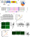
Concurrent demethylation of K450 and K451 enhances EHMT1 oncogenic activity. A, Mass spectrometry analysis on immunoprecipitated V5-EHMT1 in LNCaP cells stably overexpressing V5-tagged doxycycline-regulated EHMT1 (treated with doxycycline for 2 days) revealed methylations at K450 and K451 sites. B, The pie chart illustrating the percentage of detected unmethylated peptides (0-me), K450 or K451 methylated peptides (1-me), and K450/K451 dual-methylated peptides (2-me). C, Amino acid alignment of EHMT1 K450/451 surrounding amino acid sequence across different species and with human EHMT2 protein. D, Immunoblotting analysis of V5 in LNCaP cells stably overexpressing V5-tagged doxycycline-regulated EHMT1-WT, K450R, K451R, and K450/451R treated with doxycycline for 0–7 days. E, Proliferation assay performed on LNCaP stable cells treated with doxycycline for 1–5 days at 0.1 μg/mL. F, Transwell migration assay conducted on LNCaP stable cells expressing EHMT1 WT versus K450/451R mutant treated with or without doxycycline for 3 days. G, Immunoblotting analysis of H3K9me2 in EHMT1-K450/451R cells treated with doxycycline (for 3 days) and/or UNC0642 (0, 0.2, 2 μmol/L, for 2 days). H, Immunoblotting analysis of V5 in EHMT1-K450/451R cells treated with doxycycline (for 3 days) and/or UNC0642 (2 μmol/L, for 2 days). I, Proliferation assay performed on EHMT1-K450/451R cells treated with doxycycline and/or UNC0642 (0.2 or 2 μmol/L, for 1–4 days). J, Transwell migration assay conducted on EHMT1-K450/451R cells treated with doxycycline (for 3 days) and/or UNC0642 (2 μmol/L, for 2 days).
FIGURE 5
FIGURE 5
The dual-lysine demethylation reprograms EHMT1-mediated transcription. A, Venn diagrams showing EHMT1-WT–regulated genes versus K450/451R-regulated genes (cutoff: 2-fold, P < 0.05). B, Heat map view presenting K450/451R-regulated genes (cutoff: 2-fold, P < 0.05). C, GSEA conducted to assess the enrichment of genes regulated by the WT and mutants using hallmark gene sets (cutoff: P < 0.05). D, GSEA of genes upregulated by the overexpression of WT or K450/451R mutant in the stable lines compared with those downregulated by siEHMT1 or siEHMT2 (cutoff: P < 0.05). E, GSEA of genes upregulated by the K450/451R mutant compared with genes downregulated by siEHMT1, siEHMT2, or siLSD1 (cutoff: P < 0.05). F, Box plots depicting the relative expression of previously identified common E2F/Rb targets (49-gene) in EHMT1-K450/451R expressing LNCaP cells. G, Luciferase reporter assay measuring MCM7-promoter (E2F1 target) activity in COS7 cells transfected with E2F1 and/or EHMT1-WT or the K450/451R mutant. H, Box plots representing the doxycycline-induced change of K450/451R-upregulated or -downregulated genes in EHMT1-K450/451R stable cells (pretreated with doxycycline) treated with or without UNC0642 (2 μmol/L, for 2 days; based on RNA-seq analysis).
FIGURE 6
FIGURE 6
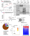
The dual-lysine demethylation dramatically expands EHMT1 chromatin occupation. A, ChIP-seq analyses of V5 were conducted on LNCaP stable cells overexpressing EHMT1 WT, K450R, K451R, and K450/451R (doxycycline pretreated for 2 days). The Venn diagram displays the overlapped sites. B, Heat map views presenting ChIP-seq signal intensity of V5 at total V5-EHMT1 binding sites. C, Chromatin distribution of EHMT1-K450/451R mutant binding sites. D, Heat map views presenting ChIP-seq signal intensity of H3K9me2 (obtained from ChIP-H3K9me2) at total V5-EHMT1 binding sites. E, ChIP-qPCR conducted for V5 binding (EHMT1 WT or K450/451R mutant) and H3K9me2 levels at two identified mutant binding sites in the stable cell lines. F, Motif enrichment analysis performed for ChIP-V5 sites identified in K450/451R mutant expressing cells.
FIGURE 7
FIGURE 7
EHMT1 can be partly demethylated by LSD1. In vitro demethylation assay measuring formaldehyde production using synthetic K450- and K451-methylated EHMT1 peptides (amino acids 441–461; A) or dual-methylated EHMT1 peptide and K4-dimethylated H3 recombinant protein (B), as substrates incubated with recombinant LSD1 protein. Results were normalized to the unmethylated EHMT1 peptide. C, Immunoblotting analysis of the indicated proteins co-immunoprecipitated with V5 or FLAG in HEK293 cells cotransfected with V5-EHMT1 WT (doxycycline for 3 days) and mutants with FLAG-LSD1. D, The Venn diagram illustrating ChIP-EHMT1 peaks in parental LNCaP cells compared with ChIP-V5 peaks in LNCaP cells expressing EHMT1-K450/451R. E, Chromatin distribution of EHMT1 binding sites was displayed. F, Motif enrichment analysis for EHMT1 binding sites. G, Heat map view presenting the ChIP-seq signal intensity of EHMT1, H3K4me2, H3K27ac, and LSD1 centered at EHMT1 binding sites in LNCaP cells.
FIGURE 8
FIGURE 8
A working model for the transcriptional repressor and activator functions of EHMT1. A graphic model illustrates how lysine demethylation enables the oncogenic activity of EHMT1.

References

    1. Yuan X, Cai C, Chen S, Chen S, Yu Z, Balk SP. Androgen receptor functions in castration-resistant prostate cancer and mechanisms of resistance to new agents targeting the androgen axis. Oncogene 2014;33:2815–25. - PMC - PubMed
    1. de Bono JS, Logothetis CJ, Molina A, Fizazi K, North S, Chu L, et al. . Abiraterone and increased survival in metastatic prostate cancer. N Engl J Med 2011;364:1995–2005. - PMC - PubMed
    1. Scher HI, Fizazi K, Saad F, Taplin ME, Sternberg CN, Miller K, et al. . Increased survival with enzalutamide in prostate cancer after chemotherapy. N Engl J Med 2012;367:1187–97. - PubMed
    1. Smith MR, Hussain M, Saad F, Fizazi K, Sternberg CN, Crawford ED, et al. . Darolutamide and survival in metastatic, hormone-sensitive prostate cancer. N Engl J Med 2022;386:1132–42. - PMC - PubMed
    1. Ge R, Wang Z, Montironi R, Jiang Z, Cheng M, Santoni M, et al. . Epigenetic modulations and lineage plasticity in advanced prostate cancer. Ann Oncol 2020;31:470–9. - PubMed

Publication types