Case Reports
. 2023 Aug 17:32:101913.
doi: 10.1016/j.ajoc.2023.101913. eCollection 2023 Dec.

Acute macular infarctions in pediatric patients with hemoglobin SS disease

Affiliations
Case Reports

Acute macular infarctions in pediatric patients with hemoglobin SS disease

Patrick Sy Lee et al. Am J Ophthalmol Case Rep. .

Abstract

Purpose: To report two cases of symptomatic posterior pole arterial occlusions in patients with hemoglobin SS disease.

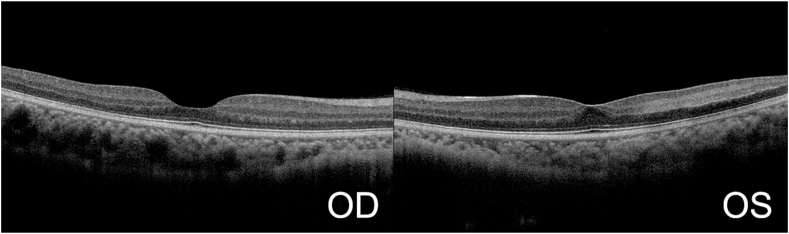
Observations: Two teenage patients with hemoglobin SS disease presented with visual distortions, and on dilated fundus examination and testing, they were found to have arterial occlusions involving the posterior pole. The patients were evaluated for stroke with head imaging and received exchange transfusion by hematology.

Conclusions and importance: This case series reports the unusual findings of arterial occlusions in the posterior pole resulting in areas of retinal whitening and ischemia in patients with HbSS. While sickle cell retinopathy is typically considered a peripheral retinal disease, these findings underscore the importance of vigilance when examining patients with sickle cell disease.

Keywords: Hemoglobin S; Ischemia; Macula; Sickle cell disease; Sickle cell retinopathy.


Conflict of interest statement

The authors declare that they have no known competing financial interests or personal relationships that could have appeared to influence the work reported in this paper.

Figures

Fig. 1
Fig. 1
Fundus photography in patient 1 showing multiple foci of whitening involving macula bilaterally.
Fig. 2
Fig. 2
Fluorescein angiography in patient 1 showing multiple areas of peripheral arterial occlusion with vascular dropout bilaterally.
Fig. 3
Fig. 3
OCT of macula in patient 1 showing significant temporal thinning of inner retinal layers, particularly pronounced in left eye.
Fig. 4
Fig. 4
Fundus photography in patient 2 showing area of whitening inferotemporally to macula in left eye.
Fig. 5
Fig. 5
Fluorescein angiography in patient 2 showing multiple areas of peripheral arterial occlusion with vascular dropout bilaterally. Note focal area of nonperfusion corresponding to area of whitening in Fig. 4.
Fig. 6
Fig. 6
OCT of macula in patient 2 showing whitening of inner retinal layers temporal to macula in left eye.

References

    1. Abdalla Elsayed M.E.A., Mura M., Al Dhibi H., et al. Sickle cell retinopathy. A focused review. Graefes Arch Clin Exp Ophthalmol. Jul 2019;257(7):1353–1364. doi: 10.1007/s00417-019-04294-2. - DOI - PubMed
    1. Gualandro S.F., Fonseca G.H., Yokomizo I.K., Gualandro D.M., Suganuma L.M. Cohort study of adult patients with haemoglobin SC disease: clinical characteristics and predictors of mortality. Br J Haematol. Nov 2015;171(4):631–637. doi: 10.1111/bjh.13625. - DOI - PubMed
    1. Nagel R.L., Fabry M.E., Steinberg M.H. The paradox of hemoglobin SC disease. Blood Rev. Sep 2003;17(3):167–178. doi: 10.1016/s0268-960x(03)00003-1. - DOI - PubMed
    1. Fox P.D., Dunn D.T., Morris J.S., Serjeant G.R. Risk factors for proliferative sickle retinopathy. Br J Ophthalmol. Mar 1990;74(3):172–176. doi: 10.1136/bjo.74.3.172. - DOI - PMC - PubMed
    1. Jin J., Miller R., Salvin J., et al. Funduscopic examination and SD-OCT in detecting sickle cell retinopathy among pediatric patients. J AAPOS. Jun 2018;22(3):197–201 e1. doi: 10.1016/j.jaapos.2017.12.019. - DOI - PubMed

Publication types

LinkOut - more resources