Review
. 2023 Oct;13(5):e200182.
doi: 10.1212/CPJ.0000000000200182. Epub 2023 Aug 30.

Differential Diagnosis of Tumor-like Brain Lesions

Affiliations
Review

Differential Diagnosis of Tumor-like Brain Lesions

Gina S Perez Giraldo et al. Neurol Clin Pract. 2023 Oct.

Abstract

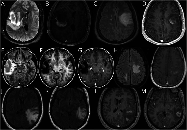
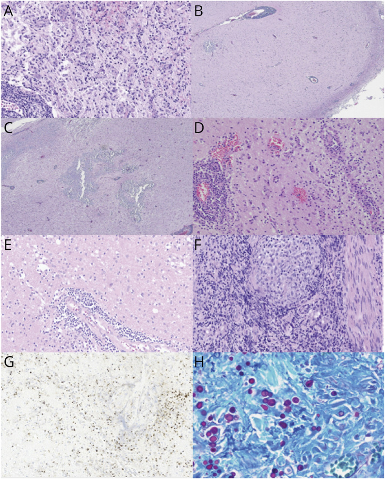
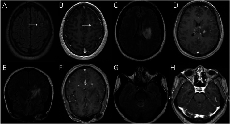
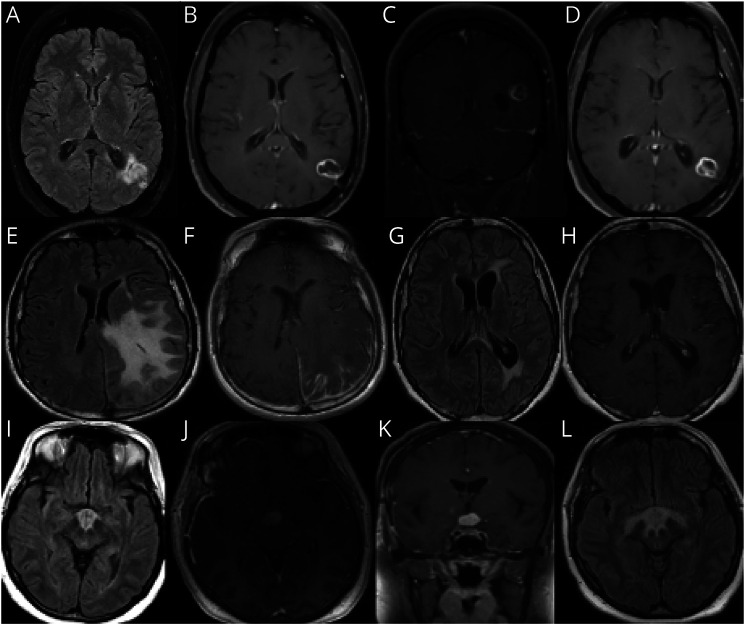
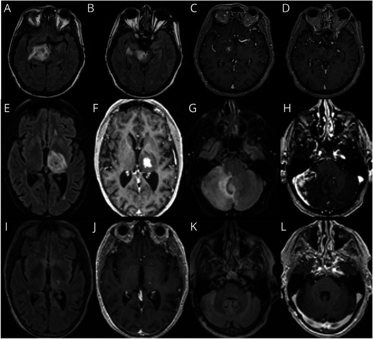
Purpose of review: Tumor-like brain lesions are rare and commonly suggest a neoplastic etiology. Failure to rapidly identify non-neoplastic causes can lead to increased morbidity and mortality. In this review, we describe 10 patients who presented with atypical, non-neoplastic tumor-like brain lesions in which brain biopsy was essential for a correct diagnosis and treatment.

Recent findings: There has been increasing recognition of autoimmune conditions affecting the nervous system, and many of those diseases can cause tumor-like brain lesions. Currently available reports of non-neoplastic tumor-like brain lesions are scarce. Most case series focus on tumefactive demyelinating lesions, and a comprehensive review including other neuroimmunological conditions such as CNS vasculitis, neurosarcoidosis, histiocytic and infectious etiologies is lacking.

Summary: We review the literature on tumor-like brain lesions intending to increase the awareness and differential diagnosis of non-neoplastic brain tumor mimics. We advocate for earlier brain biopsies, which, in our case series, significantly changed diagnosis, management, and outcomes.


Conflict of interest statement

G.S. Perez reports no disclosures relevant to the manuscript; L. Singer reports no disclosures relevant to the manuscript; T. Cao reports no disclosures relevant to the manuscript; P. Jamshidi reports no disclosures relevant to the manuscript; K. Dixit reports no disclosures relevant to the manuscript; M. Kontzialis reports no disclosures relevant to the manuscript; R. Castellani reports no disclosures relevant to the manuscript; P. Pytel reports no disclosures relevant to the manuscript; N. Anadani reports no disclosures relevant to the manuscript; C. Bevan has participated in an ad board for Genentech; E. Grebenciucova reports no disclosures relevant to the manuscript; R. Balabanov received honoraria from Biogen, Sanofi, Alexion, and Teva Pharmaceutical and research support from the National Multiple Sclerosis Society, National Institute of Health, Nextcure, and Biogen; B. Cohen reports no disclosures relevant to the manuscript. E. Graham received consulting and advisory board fees from Novartis, Atara Biotherapeutics, Tavistock Life Sciences, Horizon Therapeutics, Roche Genentech. She receives research support from F. Hoffman-La Roche Ltd. She received compensation for question writing from ACP MKSAP. Full disclosure form information provided by the authors is available with the full text of this article at Neurology.org/cp.

Figures

Figure 1
Figure 1. Demyelinating Diseases
(A–D) Patient 1, tumefactive multiple sclerosis. (E–I) Patient 2, neuromyelitis optica. (J–M) Patient 3, myelin oligodendrocyte glycoprotein antibody–associated disease (MOGAD).
Figure 2
Figure 2. Examples of Pathologic Findings
(A) Shows typical histopathologic features of a demyelinating lesion, with perivascular lymphocytes (lower left), reactive astrocytosis, and macrophage accumulation, in viable neuropil (relative axonal preservation) (magnification = ×20 before enlargement). (B, C) Images correspond to patient 2. Shows dense perivascular lymphocytic infiltrates in the meninges and the cortex, geographic areas of dystrophic calcifications and gliosis in the cortex and degenerative change in the white matter with macrophages aggregates and cavitary lesion. (D) Demonstrates histopathologic features of vasculitis, with transmural and perivascular infiltrates of mononuclear inflammatory cells affecting the medium-sized and small sized vessels (magnification = ×20 before enlargement). (E) Displays features of vasculitis with perivascular infiltrates of mononuclear inflammatory cells and intraparenchymal macrophages (magnification = ×20 before enlargement). (F) Shows features of neurosarcoidosis with well-formed non-necrotizing granuloma composed of epithelioid histiocytes (magnification = 20X before enlargement). (G) Images correspond to patient 9. Immunohistochemical stain using antibodies to Toxoplasma gondii, demonstrating tachyzoites in cerebral toxoplasmosis (magnifications = 60X before enlargement). (H) Images correspond to patient 10. Periodic acid-Schiff (PAS) special stain highlights numerous yeast-form fungal organisms with broad-based budding, demonstrating cerebral blastomycosis (magnifications = 40X before enlargement).
Figure 3
Figure 3. CNS Vasculitis
(A, B) Patient 4. (C–H) Patient 5.
Figure 4
Figure 4. Granulomatous, Histiocytic, and Plasma Cell Disorders
(A–D) Patient 6, neurosarcoidosis. (E–H) Patient 7, IgG-4–related disease (IGG4RD). (I–L) Patient 8, Langerhans histiocytosis.
Figure 5
Figure 5. Infections
(A–D) Patient 9, toxoplasmosis. (E–L) Patient 10, blastomycosis.

Similar articles

Cited by

References

    1. Frederick MC, Cameron MH. Tumefactive demyelinating lesions in multiple sclerosis and associated disorders. Curr Neurol Neurosci Rep. 2016;16(3):1-7. doi:10.1007/s11910-016-0626-9 - DOI - PubMed
    1. Algahtani H, Shirah B, Alassiri A. Tumefactive demyelinating lesions: a comprehensive review. Mult Scler Relat Disord. 2017;14:72-79. doi:10.1016/j.msard.2017.04.003 - DOI - PubMed
    1. Lucchinetti CF, Gavrilova RH, Metz I, et al. . Clinical and radiographic spectrum of pathologically confirmed tumefactive multiple sclerosis. Brain. 2008;131(7):1759-1775. doi:10.1093/brain/awn098 - DOI - PMC - PubMed
    1. Nakayama M, Naganawa S, Ouyang M, et al. . A review of clinical and imaging findings in tumefactive demyelination. Am J Roentgenol. 2021;217(1):186-197. doi:10.2214/AJR.20.23226 - DOI - PubMed
    1. Matsuoka T, Suzuki SO, Iwaki T, Tabira T, Ordinario AT, Kira JI. Aquaporin-4 astrocytopathy in Baló’s disease. Acta Neuropathol. 2010;120(5):651-660. doi:10.1007/s00401-010-0733-7 - DOI - PubMed

LinkOut - more resources