Evaluation of dual potentiality of 2,4,5-trisubstituted oxazole derivatives as aquaporin-4 inhibitors and anti-inflammatory agents in lung cells
- PMID: 37664213
- PMCID: PMC10472800
- DOI: 10.1039/d3ra03989g
Evaluation of dual potentiality of 2,4,5-trisubstituted oxazole derivatives as aquaporin-4 inhibitors and anti-inflammatory agents in lung cells
Abstract
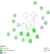
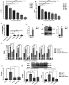
Inflammation is a multifaceted "second-line" adaptive defense mechanism triggered by exo/endogenous threating stimuli and inter-communicated by various inflammatory key players. Unresolved or dysregulated inflammation in lungs results in manifestation of diseases and leads to irreparable damage. Aquaporins (AQPs) are a ubiquitously expressed superfamily of intrinsic transmembrane water channel proteins that modulate the fluid homeostasis. In addition to their conventional functions, AQPs have clinical relevance to inflammation prevailing under the infectious conditions of various lung diseases and this proclaims them as appropriate biomarkers to be targeted. Hence an endeavor was undertaken to identify potential ligands to target AQP4 for the treatment of lung diseases. Oxazole being a versatile bio-potent core, a series of 2,4,5-trisubstituted oxazoles 3a-j were synthesized by a Lewis acid mediated reaction of aroylmethylidene malonates with nitriles. In silico studies conducted using the protein data bank (PDB) structure 3gd8 for AQP4 revealed that compound 3a would serve as a suitable candidate to inhibit AQP4 in human lung cells (NCI-H460). Further, in vitro studies demonstrated that compound 3a could effectively inhibit AQP4 and inflammatory cytokines in lung cells and hence it may be considered as a viable drug candidate for the treatment of various lung diseases.
This journal is © The Royal Society of Chemistry.
Conflict of interest statement
There are no conflicts to declare.
Figures


Similar articles
-
Aquaporins in lung health and disease: Emerging roles, regulation, and clinical implications.Respir Med. 2020 Nov-Dec;174:106193. doi: 10.1016/j.rmed.2020.106193. Epub 2020 Oct 17. Respir Med. 2020. PMID: 33096317 Free PMC article. Review.
-
Aquaporins in Respiratory System.Adv Exp Med Biol. 2023;1398:137-144. doi: 10.1007/978-981-19-7415-1_9. Adv Exp Med Biol. 2023. PMID: 36717491
-
1,3-propanediol binds deep inside the channel to inhibit water permeation through aquaporins.Protein Sci. 2016 Feb;25(2):433-41. doi: 10.1002/pro.2832. Protein Sci. 2016. PMID: 26481430 Free PMC article.
-
Aquaporin water channels: New perspectives on the potential role in inflammation.Adv Protein Chem Struct Biol. 2019;116:311-345. doi: 10.1016/bs.apcsb.2018.11.010. Epub 2019 Jan 5. Adv Protein Chem Struct Biol. 2019. PMID: 31036295 Review.
-
Role of Aquaporins in Inflammation-a Scientific Curation.Inflammation. 2020 Oct;43(5):1599-1610. doi: 10.1007/s10753-020-01247-4. Inflammation. 2020. PMID: 32435911 Review.
References
-
- Moldoveanu B. Otmishi P. Jani P. Walker J. Sarmiento X. Guardiola J. Saad M. Yu J. J. Inflammation Res. 2009;2:1–11. - PMC - PubMed
- Antonelli M. Kushner I. FASEF J. 2017;31:1787–1797. - PubMed
- Netea M.-G. Balkwill F. Chonchol M. Cominelli F. Donath M.-Y. Bourboulis G. Golenbock D. Gresnigt M.-S. Heneka M.-T. Hoffman H.-M. Hotchkiss R. Joosten A.-B. Kastner D.-L. Korte M. Latz E. Libby P. Poulsen T.-M. Mantovani A. Mills H.-G. Nowak K.-L. O'Neill L.-A. Pickkers P. Poll T.-V. Ridker P.-M. Schalkwijk J. Schwartz D.-A. Siegmund B. Steer C.-J. Tilg H. Meer W.-M. Veerdonk F.-L. Dinarello C.-A. Nat. Immunol. 2017;18:826–832. - PMC - PubMed
- Germolec D.-R. Shipkowski K.-A. Frawley R.-P. Evans E. Methods Mol. Biol. 2018;1803:57–79. - PubMed
- Zhang J.-M. An J. Int. Anesthesiol. Clin. 2007;45:27–37. - PMC - PubMed
-
- Benga G. Mol. Aspects Med. 2012;33:514–517. - PubMed
-
- Papadapoulos M.-C. Saadoun S. Verkman A. S. Pflügers Archiv: European Journal of Physiology. 2008;456:693–700. - PMC - PubMed
- Papadopoulos M.-C. Saadoun S. Biochimica et Biophysica. 2015;1848:2576–2583. - PubMed
- Edamana S. Login F.-H. Yamada S. Kwon T.-H. Nejsum L.-N. Am. J. Physiol.: Cell Physiol. 2021;320:771–777. - PubMed
LinkOut - more resources
Full Text Sources

