

Clinical and radiological characteristics of pediatric COVID-19 before and after the Omicron outbreak: a multi-center study
- PMID: 37664548
- PMCID: PMC10470622
- DOI: 10.3389/fped.2023.1172111
Clinical and radiological characteristics of pediatric COVID-19 before and after the Omicron outbreak: a multi-center study
Abstract
Introduction: The emergence of the Omicron variant has seen changes in the clinical and radiological presentations of COVID-19 in pediatric patients. We sought to compare these features between patients infected in the early phase of the pandemic and those during the Omicron outbreak.
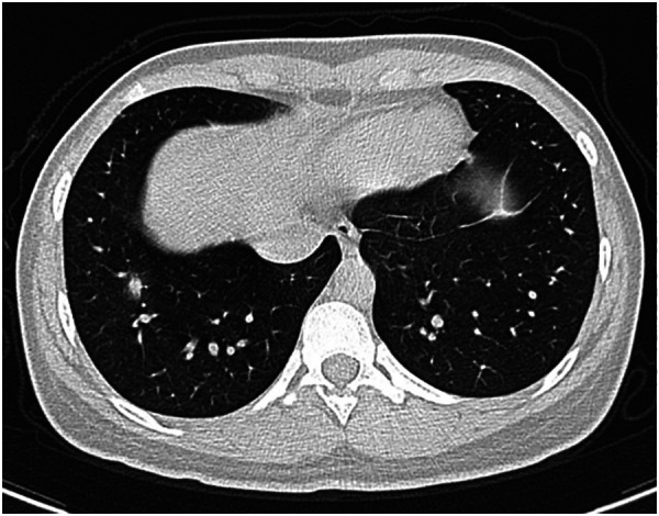
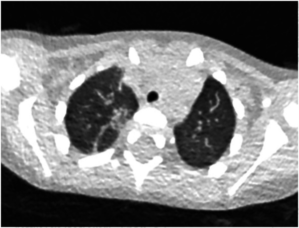
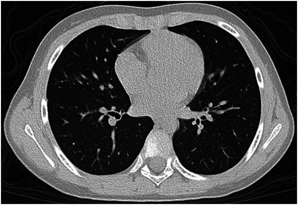
Methods: A retrospective study was conducted on 68 pediatric COVID-19 patients, of which 31 were infected with the original SARS-CoV-2 strain (original group) and 37 with the Omicron variant (Omicron group). Clinical symptoms and chest CT scans were examined to assess clinical characteristics, and the extent and severity of lung involvement.
Results: Pediatric COVID-19 patients predominantly had normal or mild chest CT findings. The Omicron group demonstrated a significantly reduced CT severity score than the original group. Ground-glass opacities were the prevalent radiological findings in both sets. The Omicron group presented with fewer symptoms, had milder clinical manifestations, and recovered faster than the original group.
Discussion: The clinical and radiological characteristics of pediatric COVID-19 patients have evolved with the advent of the Omicron variant. For children displaying severe symptoms warranting CT examinations, it is crucial to weigh the implications of ionizing radiation and employ customized scanning protocols and protective measures. This research offers insights into the shifting disease spectrum, aiding in the effective diagnosis and treatment of pediatric COVID-19 patients.
Keywords: COVID-19; CT; children; pediatric; radiation protection.
© 2023 Lin, Xu, Shen, Huang, Gao, Liu, Xie, Zhao, Xia, Lv, Ren, Zheng, Wang, Hu, Ruan and Zhang.
Conflict of interest statement
The authors declare that the research was conducted in the absence of any commercial or financial relationships that could be construed as a potential conflict of interest.
Figures


Similar articles
-
Comparative Analysis of Clinical and CT Findings in Patients with SARS-CoV-2 Original Strain, Delta and Omicron Variants.Biomedicines. 2023 Mar 14;11(3):901. doi: 10.3390/biomedicines11030901. Biomedicines. 2023. PMID: 36979880 Free PMC article.
-
Comparison of Computed Tomography and Clinical Features Between Patients Infected with the SARS-CoV-2 Omicron Variant and the Original Strain.Infect Drug Resist. 2024 Mar 5;17:807-818. doi: 10.2147/IDR.S448713. eCollection 2024. Infect Drug Resist. 2024. PMID: 38476766 Free PMC article.
-
Clinical and Pulmonary CT Characteristics of Patients Infected With the SARS-CoV-2 Omicron Variant Compared With Those of Patients Infected With the Alpha Viral Strain.Front Public Health. 2022 Jul 12;10:931480. doi: 10.3389/fpubh.2022.931480. eCollection 2022. Front Public Health. 2022. PMID: 35903393 Free PMC article.
-
SARS-CoV-2 Omicron (B.1.1.529) Variant: A Challenge with COVID-19.Diagnostics (Basel). 2023 Feb 2;13(3):559. doi: 10.3390/diagnostics13030559. Diagnostics (Basel). 2023. PMID: 36766664 Free PMC article. Review.
-
Imaging Findings of SARS-CoV-2 Infection in Pediatrics: A Systematic Review of Coronavirus Disease 2019 (COVID-19) in 850 Patients.Acad Radiol. 2020 Nov;27(11):1608-1621. doi: 10.1016/j.acra.2020.07.031. Epub 2020 Jul 30. Acad Radiol. 2020. PMID: 32773328 Free PMC article.
Cited by
-
Differences in chest imaging between Omicron and non-Omicron coronavirus disease 2019 (COVID-19) patients: a systematic review and meta-analysis.BMC Infect Dis. 2025 Apr 29;25(1):631. doi: 10.1186/s12879-025-11032-z. BMC Infect Dis. 2025. PMID: 40301746 Free PMC article.
-
Long COVID in pediatric age: an observational, prospective, longitudinal, multicenter study in Italy.Front Immunol. 2025 Apr 9;16:1466201. doi: 10.3389/fimmu.2025.1466201. eCollection 2025. Front Immunol. 2025. PMID: 40270969 Free PMC article.
References
-
- Malahe SRK, Hoek RAS, Dalm V, Broers AEC, den Hoed CM, Manintveld OC, et al. Clinical characteristics and outcomes of immunocompromised patients with coronavirus disease 2019 caused by the Omicron variant: a prospective, observational study. Clin Infect Dis. (2023) 76(3):e172–8. 10.1093/cid/ciac571 - DOI - PMC - PubMed
LinkOut - more resources
Full Text Sources
Miscellaneous