. 2023 Apr 3;16(9):1521-1533.
doi: 10.1093/ckj/sfad071. eCollection 2023 Sep.

Kidney injury molecule 1 (KIM-1): a potential biomarker of acute kidney injury and tubulointerstitial injury in patients with ANCA-glomerulonephritis

Affiliations

Kidney injury molecule 1 (KIM-1): a potential biomarker of acute kidney injury and tubulointerstitial injury in patients with ANCA-glomerulonephritis

Benoît Brilland et al. Clin Kidney J. .

Abstract

Background: Kidney injury molecule 1 (KIM-1) is a transmembrane glycoprotein expressed by proximal tubular cells, recognized as an early, sensitive and specific urinary biomarker for kidney injury. Blood KIM-1 was recently associated with the severity of acute and chronic kidney damage but its value in antineutrophil cytoplasmic antibodies (ANCA)-associated vasculitis with glomerulonephritis (ANCA-GN) has not been studied. Thus, we analyzed its expression at ANCA-GN diagnosis and its relationship with clinical presentation, kidney histopathology and early outcomes.

Methods: We assessed KIM-1 levels and other pro-inflammatory molecules (C-reactive protein, interleukin-6, tumor necrosis factor α, monocyte chemoattractant protein-1 and pentraxin 3) at ANCA-GN diagnosis and after 6 months in patients included in the Maine-Anjou registry, which gathers data patients from four French Nephrology Centers diagnosed since January 2000.

Results: Blood KIM-1 levels were assessed in 54 patients. Levels were elevated at diagnosis and decreased after induction remission therapy. KIM-1 was associated with the severity of renal injury at diagnosis and the need for kidney replacement therapy. In opposition to other pro-inflammatory molecules, KIM-1 correlated with the amount of acute tubular necrosis and interstitial fibrosis/tubular atrophy (IF/TA) on kidney biopsy, but not with interstitial infiltrate or with glomerular involvement. In multivariable analysis, elevated KIM-1 predicted initial estimated glomerular filtration rate (β = -19, 95% CI -31, -7.6, P = .002).

Conclusion: KIM-1 appears as a potential biomarker for acute kidney injury and for tubulointerstitial injury in ANCA-GN. Whether KIM-1 is only a surrogate marker or is a key immune player in ANCA-GN pathogenesis remain to be determined.

Keywords: ANCA; KIM-1; glomerulonephritis; tubulointerstitial injury; vasculitis.


Conflict of interest statement

S.W. and G.B.P. are members of the CKJ editorial board. The other authors declare that they have no conflicts of interest in relation to this article.

Figures
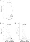
Figure 1:
Figure 1:
KIM-1 levels in ANCA-GN. (A) KIM-1 levels between ANCA-GN patients and HC. (B) KIM-1 levels at the time of ANCA-GN diagnosis and after induction remission treatment (6 months) (after exclusion of patients without KIM-1 levels assessment at 6 months). (C) KIM-1 levels at the time of ANCA-GN diagnosis and after induction remission treatment (6 months) (after exclusion of patients without KIM-1 levels detectable at diagnosis, and without KIM-1 levels assessment at 6 months).
Figure 2:
Figure 2:
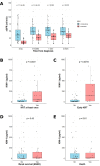
KIM-1 levels according to relevant outcomes. (A) Evolution of eGFR overtime. (B) Need for KRT during follow-up. (C) Need for early KRT (within 30 days from diagnosis). (D) Renal survival (ESKD). (E) Patient survival (death).
Figure 3:
Figure 3:
Correlation between KIM-1 levels and ANCA-GN presentation. Correlation has been assessed with Spearman method. Correlation coefficient ranges from –1 (dark red) to +1 (dark blue). *P-value <.05. “% normal glom.,” “% crescentic glom.” and “% sclerotic glom.” refer to the amount for glomerular involvement on kidney biopsy. IFTA: interstitial fibrosis/tubular atrophy on kidney biopsy.
Figure 4:
Figure 4:
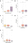
KIM-1 levels and histological lesions found on kidney biopsy. KIM-1 levels according to the severity of ATN (A), the amount of IF/TA (B), the severity of interstitial infiltrate (C), Berden classification (D) and Renal Risk Score (E). IF/TA was scored quantitatively as in the Banff classification [31], and trichotomized in three classes: ≤5%, 5%–25%, >25%. ATN was scored as previously defined [32] (n1 = loss of brush border, n2 = tubular epithelial cells in the lumen, vacuolization of tubular cells, n3 = exposure of basal membranes/epithelial necrosis). Interstitial infiltrate was scored semi-quantitatively as in Banff classification [31] (i1: ≤25%, i2: 25%–50%, i3: ≥50%), without i0 stage because no patients were found with the absence of infiltration.

References

    1. Kitching AR, Anders H-J, Basu Net al. . ANCA-associated vasculitis. Nat Rev Dis Primers 2020;6:71. 10.1038/s41572-020-0204-y. - DOI - PubMed
    1. Sinico RA, Di Toma L, Radice A.. Renal involvement in anti-neutrophil cytoplasmic autoantibody associated vasculitis. Autoimmun Rev 2013;12:477–82. 10.1016/j.autrev.2012.08.006. - DOI - PubMed
    1. Berden AE, Ferrario F, Hagen ECet al. . Histopathologic classification of ANCA-associated glomerulonephritis. J Am Soc Nephrol 2010;21:1628–36. 10.1681/ASN.2010050477. - DOI - PubMed
    1. van Daalen EE, Wester Trejo MAC, Göçeroğlu Aet al. . Developments in the histopathological classification of ANCA-associated glomerulonephritis. Clin J Am Soc Nephrol 2020;15:1103–11. 10.2215/CJN.14561119. - DOI - PMC - PubMed
    1. Wallace ZS, Miloslavsky EM. Management of ANCA associated vasculitis. BMJ 2020;m421. 10.1136/bmj.m421. - DOI - PubMed