. 2023 Oct 13;51(18):10049-10058.
doi: 10.1093/nar/gkad724.

Structural basis of transcriptional activation by the OmpR/PhoB-family response regulator PmrA

Affiliations

Structural basis of transcriptional activation by the OmpR/PhoB-family response regulator PmrA

Yuan-Chao Lou et al. Nucleic Acids Res. .

Abstract

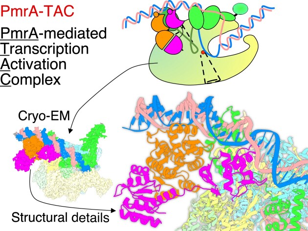
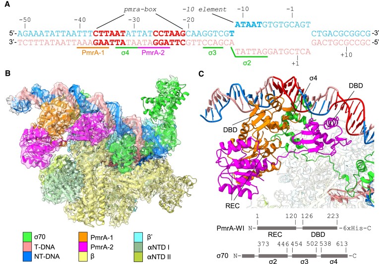
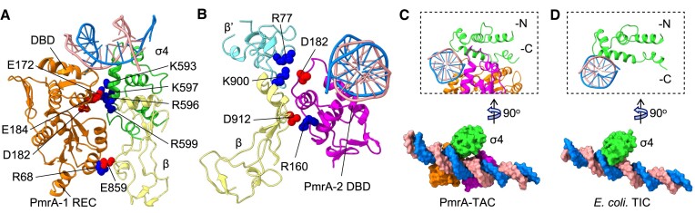
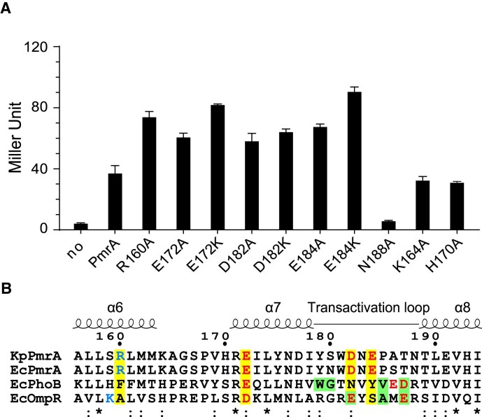
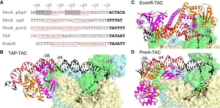
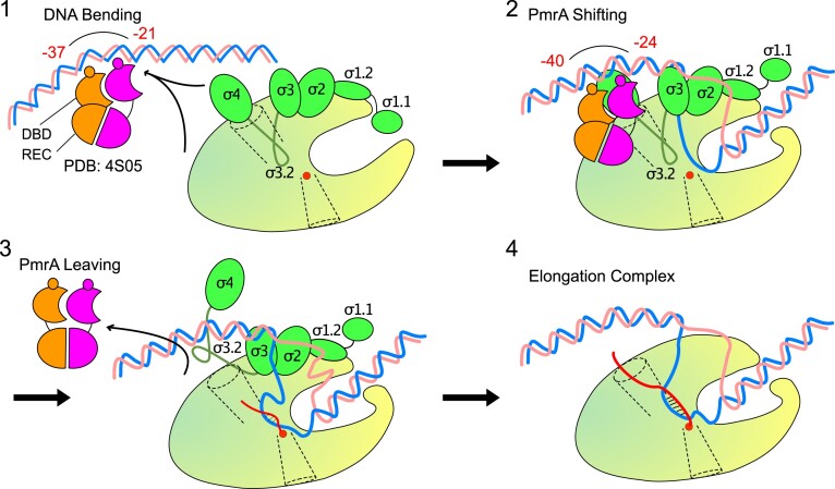
PmrA, an OmpR/PhoB-family response regulator, triggers gene transcription responsible for polymyxin resistance in bacteria by recognizing promoters where the canonical-35 element is replaced by the pmra-box, representing the PmrA recognition sequence. Here, we report a cryo-electron microscopy (cryo-EM) structure of a bacterial PmrA-dependent transcription activation complex (TAC) containing a PmrA dimer, an RNA polymerase σ70 holoenzyme (RNAPH) and the pbgP promoter DNA. Our structure reveals that the RNAPH mainly contacts the PmrA C-terminal DNA-binding domain (DBD) via electrostatic interactions and reorients the DBD three base pairs upstream of the pmra-box, resulting in a dynamic TAC conformation. In vivo assays show that the substitution of the DNA-recognition residue eliminated its transcriptional activity, while variants with altered RNAPH-interacting residues resulted in enhanced transcriptional activity. Our findings suggest that both PmrA recognition-induced DNA distortion and PmrA promoter escape play crucial roles in its transcriptional activation.


Figures

Graphical Abstract
Graphical Abstract
Figure 1.
Figure 1.
Cryo-EM structure of the PmrA-dependent TAC. (A) The synthetic promoter DNA scaffolds used in the PmrA TAC, with the pbgP promoter sequence from –62 to –13, and a transcription bubble sequence including the -10 element and discriminator from –12 to the downstream region to mimic transcription initiation. The nucleotide positions are numbered relative to the transcription start site. The template strand (T-DNA) and the non-template strand (NT-DNA) of DNA are colored in salmon and blue, respectively. Two hexanucleotide repeats in the pmra-box are colored in red. The DNA sequences associated with binding of PmrA, σ4, σ3 and σ2 are labeled and underlined. (B) A composite map containing the RNAP core and the 3.70 Å focused map (PmrA-WI, the upstream DNA and part of σ70) is overlaid with the built PmrA TAC complex structure. An EM map with partial transparency is used to present the good superposition of the map and structure. (C) PmrA-WI, the upstream DNA and part of σ70 demonstrate the interactions between PmrA and DNA. DNA regions are colored as in (A). The upstream and downstream PmrA-WI protomers are denoted as PmrA-1 and PmrA-2, and are colored in orange and magenta, respectively. The σ70 are in green. Domain architectures of PmrA and σ70 are also provided. The REC and DBD of PmrA and σ4, σ3 and σ2 of σ70 are indicated. The RNAP core proteins are uncolored.
Figure 2.
Figure 2.
Analysis of the PmrA–RNAPH interface. Dimeric PmrA-WI interacts with the double-stranded DNA, the σ4 domain of the σ70 cofactor and the RNAP core β and β′ subunits (colored as in Figure 1B). (A) Negatively charged PmrA-1 DBD residues E172, D182 and E184 form electrostatic interactions with positively charged residues K593, R596, K597 and R599 of the σ4 domain of the σ70 cofactor. The REC residue R68 is also located near E859 of the RNAP β subunit, contributing to complex stabilization. The listed residues are presented in a single-ball mode. (B) PmrA-2 DBD, which binds to the downstream DNA, interacts electrostatically with the RNAP β and β′ subunits. (C) The weaker interaction between the σ4 domain and DNA in the PmrA TAC compared with (D) the E. coli RNAP σ70 transcription initiation complex (TIC; PDB: 6CA0) due to the binding of the two PmrA DBDs to the -35 position of the DNA. A lateral view (right top corner) shows that in the PmrA TAC, the σ4 domain is not inserted as deeply into the DNA major groove as it is in the E. coli TIC without a transcription factor.
Figure 3.
Figure 3.
Upstream-shifted DBD–DNA interaction in the PmrA TAC. (A) The comparison of the PmrA DBD–DNA interactions in the TAC with the crystal structure of PmrA–DNA (PDB ID 4S05) involved superimposing the double-stranded DNA fragment from positions –38 to –20, depicted in a similar orientation to that in Figure 1C. A 90° rotation view (B) and separate depictions of the DBD–DNA structures in 4S05 (C) and in the PmrA TAC (D) provides clear visualization of their relative orientations. A lateral view (E) shows the DNA and downstream DBDs, highlighting the shifting of the DBD recognition helix in two complex structures. RNAPH is colored as in Figure 1B, with DBDs in the TAC shown in orange and magenta, and in 4S05 in dark gray and black. Residue N188 on the recognition helix is displayed as a sphere. The imperfect repeat sequences ‘CTTAAT’ and ‘CCTAAG’ in the pmra-box are emphasized in blue, their complementary sequences in salmon and other nucleotides in gray. The DNA is visualized as sticks in (C) and as rings in (D). Residue N188, the C-terminal β hairpin (Cβ) and the ‘AA’ sequences are labeled and indicated in (C) and (D).
Figure 4.
Figure 4.
Residues involved in transcriptional activation. (A) In vivo β-galactosidase reporter assay in K. pneumoniae carrying plasmids that express PmrA or its variants plus the reporter plasmid hosting the pbgP promoter in front of the lacZ gene. Expression of PmrA or its variants was induced by IPTG (1 mM/ml). Assay for PmrA without IPTG addition is labeled as ‘no’. Results are expressed as Miller Units. All experiments were performed in triplicate. Error bars represent the standard deviation. The horizontal dashed line indicates the value obtained from PmrA. (B) Sequence alignment of K. pneumoniae PmrA, and PmrA, PhoB and OmpR from E. coli. K. pneumoniae PmrA residues involved in RNAPH interactions are highlighted with a yellow background and are colored in blue and red for positive and negative charges, respectively. Residues for which replacement abolishes transcriptional activation by E. coli PhoB and OmpR are shown with a green background. Fully conserved or similar residues are denoted * or :, respectively.
Figure 5.
Figure 5.
Comparison of promoter sequences and TAC architectures. (A) Alignment of the pbgP, ugd (for PmrA), pstS (for PhoB) and promoter sequences for TAP (a T. thermophilus homolog of E. coli CAP) and EcmrR (MerR family regulator) transcription factors. The transcription factor recognition sites are marked in red and underlined. Shifted PmrA-binding sites in the PmrA TAC are highlighted with a gray background. The -10 elements are marked in bold. The structures of TAP TAC (PDB: 5I2D), EcmrR TAC (PDB: 6XL5) and PmrA TAC are shown in (B), (C) and (D), respectively. The transcription factors adopt a dimeric conformation and are colored in orange and magenta. RNAPH is colored as in Figure 1B. The DNA is colored as indicated in (A), with the -35 and -10 positions labeled.
Figure 6.
Figure 6.
Schematic illustration of four steps in PmrA-regulated transcriptional initiation. RNAP is shown as a yellow-green oval with a main channel cleft. DNA, PmrA dimer and σ70 are colored as in Figure 1B. The catalytic Mg2+ ion is displayed as a red sphere. The nascent RNA is shown as a curvy red line. The RNA exit channel is shown as a funnel in dotted lines.

Similar articles

Cited by

References

    1. Chen H.D., Groisman E.A.. The biology of the PmrA/PmrB two-component system: the major regulator of lipopolysaccharide modifications. Annu. Rev. Microbiol. 2013; 67:83–112. - PMC - PubMed
    1. Mitrophanov A.Y., Jewett M.W., Hadley T.J., Groisman E.A.. Evolution and dynamics of regulatory architectures controlling polymyxin B resistance in enteric bacteria. PLoS Genet. 2008; 4:e1000233. - PMC - PubMed
    1. Gunn J.S. The Salmonella PmrAB regulon: lipopolysaccharide modifications, antimicrobial peptide resistance and more. Trends Microbiol. 2008; 16:284–290. - PubMed
    1. Marchal K., De Keersmaecker S., Monsieurs P., van Boxel N., Lemmens K., Thijs G., Vanderleyden J., De Moor B.. In silico identification and experimental validation of PmrAB targets in Salmonella typhimurium by regulatory motif detection. Genome. Biol. 2004; 5:R9. - PMC - PubMed
    1. Cheng H.Y., Chen Y.F., Peng H.L.. Molecular characterization of the PhoPQ–PmrD–PmrAB mediated pathway regulating polymyxin B resistance in Klebsiella pneumoniae CG43. J. Biomed. Sci. 2010; 17:60. - PMC - PubMed

Publication types

MeSH terms