. 2023 Sep 15;150(18):dev201666.
doi: 10.1242/dev.201666. Epub 2023 Sep 15.

Ets-1 transcription factor regulates glial cell regeneration and function in planarians

Affiliations

Ets-1 transcription factor regulates glial cell regeneration and function in planarians

Bidushi Chandra et al. Development. .

Abstract

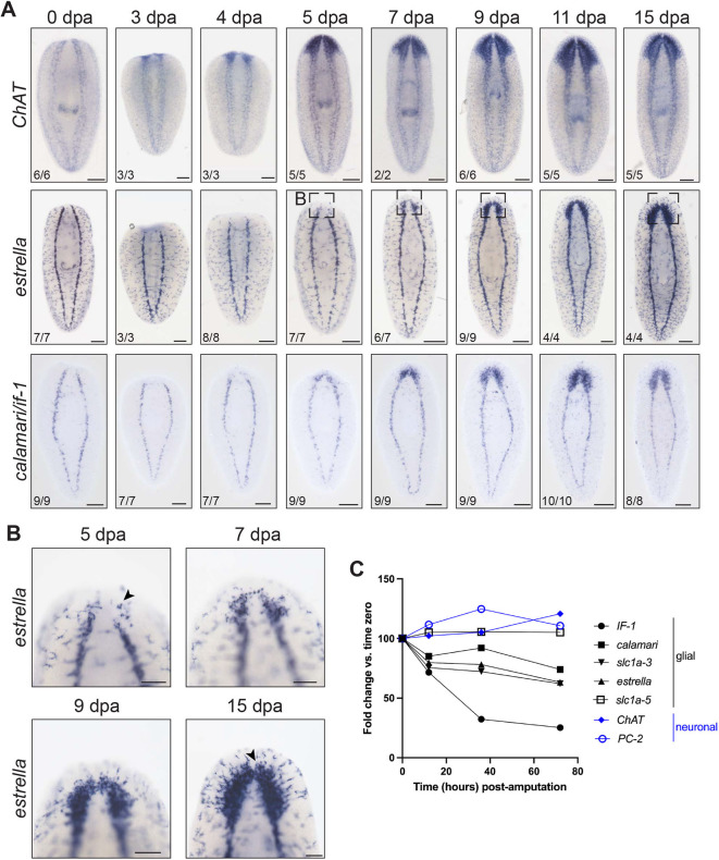
Glia play multifaceted roles in nervous systems in response to injury. Depending on the species, extent of injury and glial cell type in question, glia can help or hinder the regeneration of neurons. Studying glia in the context of successful regeneration could reveal features of pro-regenerative glia that could be exploited for new human therapies. Planarian flatworms completely regenerate their nervous systems after injury - including glia - and thus provide a strong model system for exploring glia in the context of regeneration. Here, we report that planarian glia regenerate after neurons, and that neurons are required for correct glial numbers and localization during regeneration. We also identify the planarian transcription factor-encoding gene ets-1 as a key regulator of glial cell maintenance and regeneration. Using ets-1 (RNAi) to perturb glia, we show that glial loss is associated with altered neuronal gene expression, impeded animal movement and impaired nervous system architecture - particularly within the neuropil. Importantly, our work reveals the inter-relationships of glia and neurons in the context of robust neural regeneration.

Keywords: S. mediterranea; ETS; Glia; Nervous system; Planarian; Regeneration.


Conflict of interest statement

Competing interests The authors declare no competing or financial interests.

Figures

Fig. 1.
Fig. 1.
Planarian glial cells regenerate after neurons. (A) In situ hybridization regeneration timeline of neurons (top), marked by choline acetyltransferase (ChAT), and glial cells (bottom), marked by estrella or pooled if-1/calamari. (B) Higher magnification of estrella expression in head blastema at 5, 7, 9 and 15 dpa. Arrowheads indicate round estrella+ cells (5 dpa) that progress to stellate morphology (15 dpa). (C) RNA-seq of tail fragments regenerating head tissue illustrates glial and neuronal marker transcript levels at early time points post-amputation (re-analysis of data from Roberts-Galbraith et al., 2016). Planarian glial markers are downregulated in the first 72 h post-amputation. Ventral views, anterior upwards. Scale bars: 200 µm in A; 100 µm in B.
Fig. 2.
Fig. 2.
Planarian glial cells develop after neurons during embryogenesis. (A) Single embryo RNA-sequencing shows expression of neuronal and glial markers (re-analysis of data from Davies et al., 2017). (B) In situ hybridization of neuronal (ChAT) and glial (if-1, calamari and estrella) markers in planarian embryos (early S6, mid-S6, S7, S7.5 and S8) and juveniles (1 week post-hatching); ventral views. Arrowheads indicate estrella expression in mouth (S7) and peripheral nervous system (S8). (C) In situ hybridization of ChAT and estrella on staged embryos; dorsal views. Higher magnification images show expression near and within the eyespot. estrella expression near the eye appears at S8 (arrowhead); ChAT expression, in contrast, is seen as early as S7. Anterior is leftwards. Scale bars: 200 µm; 50 µm (higher magnification images).
Fig. 3.
Fig. 3.
The presence of estrella+ cells is neuron dependent. (A) Fluorescence in situ hybridization of regenerated control, coe(RNAi) and sim(RNAi) animals, detecting ChAT (neurons, magenta) and estrella (glia, green) transcripts and stained using DAPI (nuclei, blue). (B) Quantification of brain-to-body ratio [normalized to control (100%)]. Unpaired t-test with Welch's correction. (C) Quantification of estrella+ cells in the head region in 250 µm2 areas. Unpaired t-test with Welch's correction. (D) Fluorescence in situ hybridization of regenerated control and ndk(RNAi) animals detecting ChAT (magenta) and estrella (green), and stained with DAPI (blue). (E) Quantification of normalized brain-to-body ratio. Unpaired t-test with Welch's correction. (F) Quantification of estrella+ cells in expanded posterior of the brain (depicted in illustration) in control and ndk(RNAi) animals. Unpaired t-test with Welch's correction. (G) Fluorescence in situ hybridization of regenerated control and ovo(RNAi) animals detecting pooled sans/foxQ2/myoVIIA (photoreceptor neurons, magenta) and estrella (green), and stained with DAPI (blue). Arrowheads indicate estrella+ cells in or near the eyespot. (H) Quantification of estrella+ (black) or estrella (white) eyespots after control or ovo(RNAi). Fisher's exact test. (I) Fluorescence in situ hybridization of control and soxB1-2(RNAi) animals detecting pooled pkd2L-1/pkd1L-2 (sensory neurons, magenta) and estrella (green), and stained with DAPI (blue) in medial stripe and lateral margins in non-regenerated trunk tissue (dorsal view). Quantification of pkd+ or estrella+ cells in respective regions illustrated on the left. Unpaired t-test with Welch's correction (n=18). *P≤0.05, **P≤0.01, ***P≤0.001, ****P≤0.0001; ns, not significant. Data are mean±s.d. Scale bars: 50 µm in A,G; 200 µm in D,I.
Fig. 4.
Fig. 4.
ets-1 affects glial gene expression. (A) Whole-mount in situ hybridization of ets-1 in uninjured untreated animals. (B) RNAi feeding paradigm for ets-1(RNAi). Feeding (F), amputation (A), kill/fix (K/F) and staining (S) are indicated. (C,D) In situ hybridization of ets-1(RNAi) regenerated animals detecting calamari or estrella expression. Red arrowhead indicates reduced peripheral estrella+ cells. (E) RT-qPCR used to detect levels of ets-1, if-1, estrella and calamari transcripts after RNAi and regeneration. Unpaired t-test. Data are mean±s.e.m. (F) Fluorescence in situ hybridization for calamari (magenta) and estrella (green), and stained with DAPI (cell nuclei, blue) in newly regenerated heads in control and ets-1(RNAi) animals. White arrowhead indicates cali+/estrella; black arrowhead indicated cali+/estrella+; arrow indicates cali/estrella+. (G-K) Quantification of glia markers in 250 µm2 areas within head blastemas from F. Unpaired t-test with Welch's correction. Data are mean±s.d. (L-N) Illustration showing 200 µm2 squares used to quantify PNS estrella+ cells. Images and quantifications show reduced peripheral estrella+ cells in ets-1(RNAi) animals (see also red arrowhead in D). Unpaired t-test with Welch's correction. Dashed lines indicate amputation sites. Data are mean±s.d. **P≤0.01, ***P≤0.001, ****P≤0.0001; ns, not significant. Scale bar: 200 µm in A,C,D,F; 50 µm in L.
Fig. 5.
Fig. 5.
Knockdown of ets-1 does not affect neuronal cell numbers. (A) ChAT in situ hybridization of regenerated control and ets-1(RNAi) animals. Dashed line indicates amputation site. (B) Brain-to-body ratio quantification shows reduced brain area for ets-1(RNAi) animals. Unpaired t-test with Welch's correction. Data are mean±s.d. (C) RT-qPCR was used to detect levels of ets-1 and ChAT transcripts in regenerated RNAi animals; the same cDNA samples were used for Fig. 5F,H,K,O,T. Unpaired t-test. Data are mean±s.e.m. (D) 7 dpa control and ets-1(RNAi) animals subjected to in situ hybridization for neuropeptide precursor-3 (npp3). (E) npp-3+ cells counted in 200 µm2 areas throughout the body. Unpaired t-test with Welch's correction. Data are mean±s.d. (F) npp-3 transcript levels detected with RT-qPCR in regenerated RNAi animals. Unpaired t-test. Data are mean±s.e.m. (G) In situ hybridization of 7 dpa control and ets-1(RNAi) animals showing secreted peptide prohorome-12 (spp12). (H) spp-12 transcript levels detected with RT-qPCR in regenerated RNAi animals. Unpaired t-test. Data are mean±s.e.m. (I) In situ hybridization of control and ets-1(RNAi) animals showing glutamic acid decarboxylase (gad). 62.5% of ets-1(RNAi) animals had disorganized gad+ arch pattern. (J) Quantification of gad+ cells, normalized to body size. Unpaired t-test with Welch's correction. Data are mean±s.d. (K) gad transcripts levels detected with RT-qPCR in regenerated RNAi animals. Unpaired t-test. Data are mean±s.e.m. (L) In situ hybridization of regenerated animals in control and ets-1(RNAi) animals with tryptophan hydroxylase (tph). (M,N) Quantification of tph+ cells in specific areas throughout the body and within the eye compared with body size. Unpaired t-test with Welch's correction. Data are mean±s.d. (O) RT-qPCR detecting tph transcript levels in regenerated RNAi animals. Unpaired t-test. Data are mean±s.e.m. (P) 7 dpa control and ets-1(RNAi) animals subjected to fluorescence in situ hybridization for tyrosine hydroxylase (th, magenta) and stained with DAPI (gray). (Q) Average intensity of th fluorescence in situ hybridization was quantified for control and ets-1(RNAi) animals. Unpaired t-test with Welch's correction. Data are mean±s.d. (R) th+ cells in the PNS were counted in 100 µm2 areas. Unpaired t-test with Welch's correction. Data are mean±s.d. (S) Quantification of th+ cells in the CNS was quantified in control and ets-1(RNAi) animals, and normalized to body size. Unpaired t-test with Welch's correction. Data are mean±s.d. (T) RT-qPCR detecting th transcript levels in regenerated RNAi animals. Unpaired t-test. Data are mean±s.e.m. *P≤0.05, **P≤0.01, ****P≤0.0001; ns, not significant. Scale bars: 200 µm in A,D,G,L; 100 µm in I; 50 µm in P.
Fig. 6.
Fig. 6.
ets-1 knockdown results in changes in neural architecture. (A) 7 dpa regenerated control and ets-1(RNAi) animals were subjected to immunofluorescence against arrestin (photoreceptor axons, green) and stained with DAPI (blue). ets-1(RNAi) animals exhibited several defects in axon fasciculation (arrowheads). (B) Percentage of control and ets-1(RNAi) animals exhibiting one or more defects in axon fasciculation. See Materials and Methods for our criteria for ‘irregular’ organization. Fisher's exact test. (C) Immunofluorescence for anti-synapsin (synapses, green) and DAPI staining (nuclei, blue) in regenerated control and ets-1(RNAi) animals. Dashed line indicates amputation site. (D) Illustration of neuropil width measurement. (E,F) Quantification of neuropil width (E) and average fluorescence intensity (F). Unpaired t-test with Welch's correction. (G) Immunofluorescence for anti-synapsin (green) and DAPI staining (nuclei, blue) in uninjured control and ets-1(RNAi) animals. (H) Quantification of average fluorescence intensity. Unpaired t-test with Welch's correction. *P≤0.05, **P≤0.01; ns, not significant. Data are mean±s.d. Scale bars: 20 µm in A; 100 µm in C; 200 µm in G.
Fig. 7.
Fig. 7.
ets-1 knockdown leads to changes in planarian behavior. (A) Illustration of light/dark assay (Paskin et al., 2014; Zewde et al., 2018). (B) Graph shows percentage of intact animals on the light side (n=10-12 animals per replicate; three replicates, see also Fig. S9A). (C) Quantification of animals that exhibit inch-worming in the context of a light/dark assay. Fisher's exact test. (D) In an open field, ets-1(RNAi) still led to a significantly higher incidence of inch-worming behavior. Fisher's exact test. *P≤0.05, ****P≤0.0001.

Similar articles

Cited by

References

    1. Adams, K. L. and Gallo, V. (2018). The diversity and disparity of the glial scar. Nat. Neurosci. 21, 9-15. 10.1038/s41593-017-0033-9 - DOI - PMC - PubMed
    1. Aldskogius, H. and Kozlova, E. N. (1998). Central neuron–glial and glial–glial interactions following axon injury. Prog. Neurobiol. 55, 1-26. 10.1016/S0301-0082(97)00093-2 - DOI - PubMed
    1. Alesci, A., Pergolizzi, S., Lo Cascio, P., Fumia, A. and Lauriano, E. R. (2022). Neuronal regeneration: vertebrates comparative overview and new perspectives for neurodegenerative diseases. Acta Zool. 103, 129-140. 10.1111/azo.12397 - DOI
    1. Allen, N. J. and Lyons, D. A. (2018). Glia as architects of central nervous system formation and function. Science 362, 181-185. 10.1126/science.aat0473 - DOI - PMC - PubMed
    1. Amouyel, P., Gégonne, A., Delacourte, A., Défossez, A. and Stéhelin, D. (1988). Expression of ETS proto-oncogenes in astrocytes in human cortex. Brain Res. 447, 149-153. 10.1016/0006-8993(88)90976-6 - DOI - PubMed

Publication types

Substances