Focused Shear Wave Beam Propagation in Tissue-Mimicking Phantoms
- PMID: 37665711
- PMCID: PMC10951862
- DOI: 10.1109/TBME.2023.3311688
Focused Shear Wave Beam Propagation in Tissue-Mimicking Phantoms
Abstract
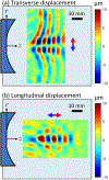
Objective: Ultrasound transient elastography (TE) technologies for liver stiffness measurement (LSM) utilize vibration of small, flat pistons, which generate shear waves that lack directivity. The most common cause for LSM failure in practice is insufficient shear wave signal at the needed depths. We propose to increase shear wave amplitude by focusing the waves into a directional beam. Here, we demonstrate the generation and propagation of focused shear wave beams (fSWBs) in gelatin.
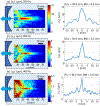
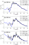
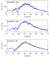
Methods: Directional fSWBs are generated by vibration at 200-400 Hz of a concave piston embedded near the surface of gelatin phantoms and measured with high-frame-rate ultrasound imaging. Five phantoms with a range of stiffnesses are employed. Shear wave speeds assessed by fSWBs are compared with those by radiation-force-based methods (2D SWE). fSWB amplitudes are compared to predictions using an analytical model.
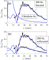
Results: fSWB-derived shear wave speeds are in good agreement with 2D SWE. The amplitudes of fSWBs are localized to the LSM region and are significantly greater than unfocused shear waves. Overall agreement with theory is observed, with some discrepancies in the theoretical source condition.
Conclusion: Focusing shear waves can increase the signal in the LSM region for TE. Challenges for translation include coupling piston vibration with the patient skin and increased attenuation in vivo compared to the phantoms employed here.
Significance: Fibrosis is the most predictive measure of patient outcome in non-alcoholic fatty liver disease. Increased shear wave amplitude in the LSM region can reduce fibrosis assessment failure rates by TE, thus reducing the need for invasive methods like biopsy.
Figures




Similar articles
-
Ultrasound Shear Wave Elastography for Liver Disease. A Critical Appraisal of the Many Actors on the Stage.Ultraschall Med. 2016 Feb;37(1):1-5. doi: 10.1055/s-0035-1567037. Epub 2016 Feb 12. Ultraschall Med. 2016. PMID: 26871407
-
Inflammatory activity affects the accuracy of liver stiffness measurement by transient elastography but not by two-dimensional shear wave elastography in non-alcoholic fatty liver disease.Liver Int. 2022 Jan;42(1):102-111. doi: 10.1111/liv.15116. Epub 2021 Dec 3. Liver Int. 2022. PMID: 34821035 Free PMC article.
-
Three-dimensional shear wave elastography on conventional ultrasound scanners with external vibration.Phys Med Biol. 2020 Nov 5;65(21):215009. doi: 10.1088/1361-6560/aba5ea. Phys Med Biol. 2020. PMID: 32663816 Free PMC article.
-
Technical performance of shear wave elastography for measuring liver stiffness in pediatric and adolescent patients: a systematic review and meta-analysis.Eur Radiol. 2019 May;29(5):2560-2572. doi: 10.1007/s00330-018-5900-6. Epub 2019 Jan 7. Eur Radiol. 2019. PMID: 30617493
-
Liver fibrosis assessment: MR and US elastography.Abdom Radiol (NY). 2022 Sep;47(9):3037-3050. doi: 10.1007/s00261-021-03269-4. Epub 2021 Oct 23. Abdom Radiol (NY). 2022. PMID: 34687329 Free PMC article. Review.
Cited by
-
Acoustic radiation force-induced longitudinal shear wave for ultrasound-based viscoelastic evaluation.Ultrasonics. 2024 Aug;142:107389. doi: 10.1016/j.ultras.2024.107389. Epub 2024 Jun 22. Ultrasonics. 2024. PMID: 38924960 Free PMC article.
-
Collagen fibers quantification for liver fibrosis assessment using linear dichroism photoacoustic microscopy.Photoacoustics. 2025 Feb 3;42:100694. doi: 10.1016/j.pacs.2025.100694. eCollection 2025 Apr. Photoacoustics. 2025. PMID: 39996157 Free PMC article.
-
Design and application of a realistic and multifunctional retinal phantom for standardizing ophthalmic imaging systems.Commun Eng. 2025 Jul 26;4(1):134. doi: 10.1038/s44172-025-00475-6. Commun Eng. 2025. PMID: 40715531 Free PMC article.
-
Integrated Difference Autocorrelation: A Novel Approach to Estimate Shear Wave Speed in the Presence of Compression Waves.IEEE Trans Biomed Eng. 2025 Feb;72(2):586-594. doi: 10.1109/TBME.2024.3464104. Epub 2025 Jan 21. IEEE Trans Biomed Eng. 2025. PMID: 39302787
-
Mechanically anisotropic phantoms for magnetic resonance elastography.Magn Reson Med. 2025 May;93(5):2123-2139. doi: 10.1002/mrm.30394. Epub 2024 Dec 3. Magn Reson Med. 2025. PMID: 39627953
References
-
- Pillai A et al. “Diagnostic accuracy of shear-wave elastography for breast lesion characterization in women: A systematic review and meta-analysis,” J. Am. Coll. Radiol 19, 625–634 (2022). - PubMed
-
- Creze M et al. “Shear wave sonoelastography of skeletal muscle: basic principles, biomechanical concepts, clinical applications, and future perspectives,” Skeletal Radiol 47, 457–471 (2018). - PubMed
Publication types
MeSH terms
Substances
Grants and funding
LinkOut - more resources
Full Text Sources

