. 2023 Sep;9(9):1500-1513.
doi: 10.1038/s41477-023-01478-x. Epub 2023 Sep 4.

Antigravitropic PIN polarization maintains non-vertical growth in lateral roots

Affiliations

Antigravitropic PIN polarization maintains non-vertical growth in lateral roots

Suruchi Roychoudhry et al. Nat Plants. 2023 Sep.

Abstract

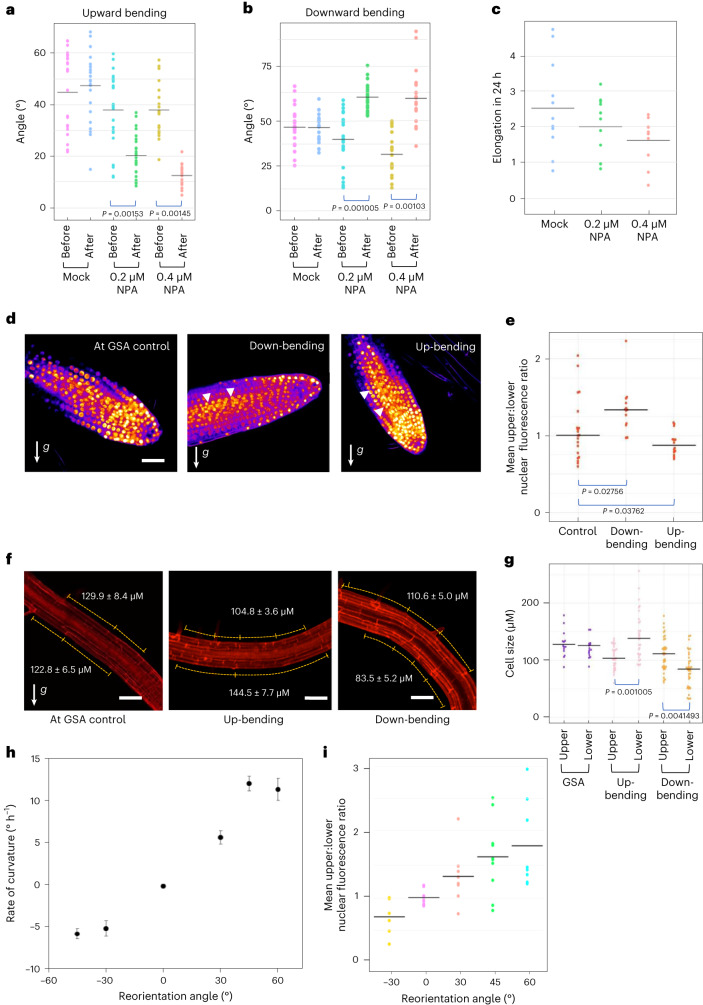
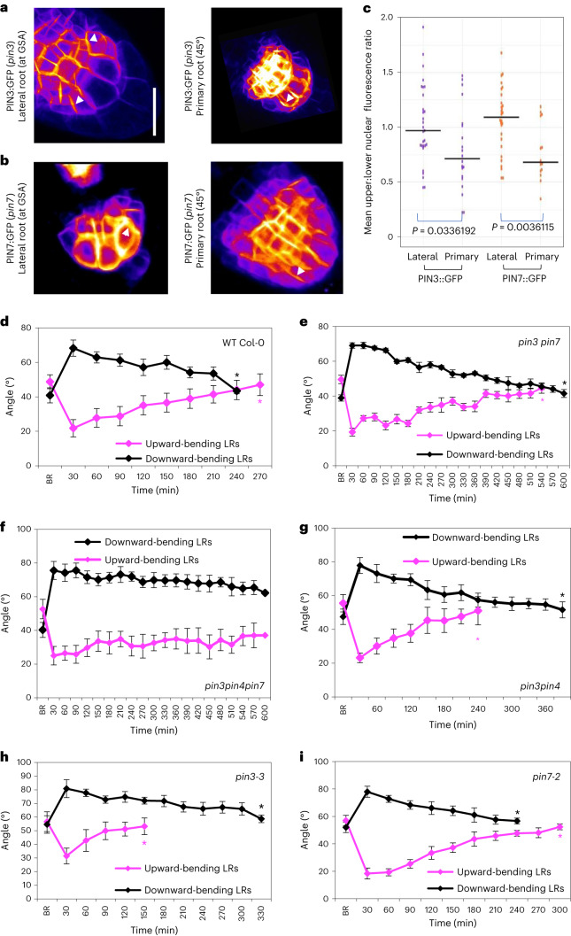
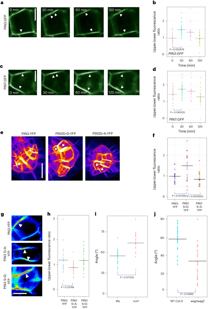
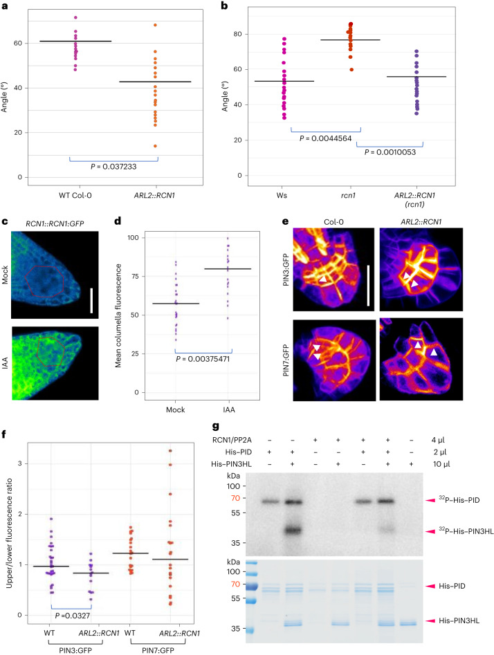
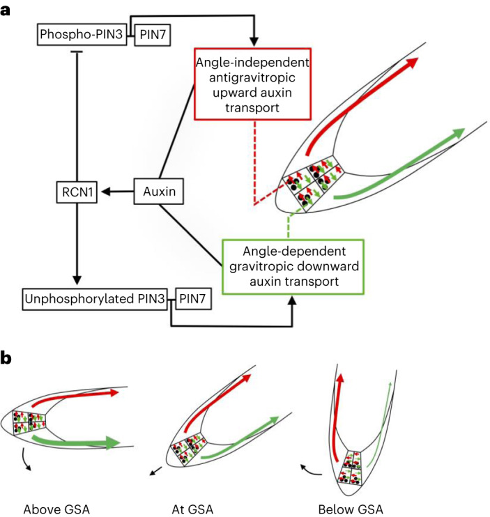
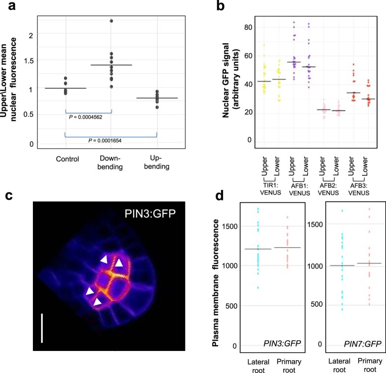
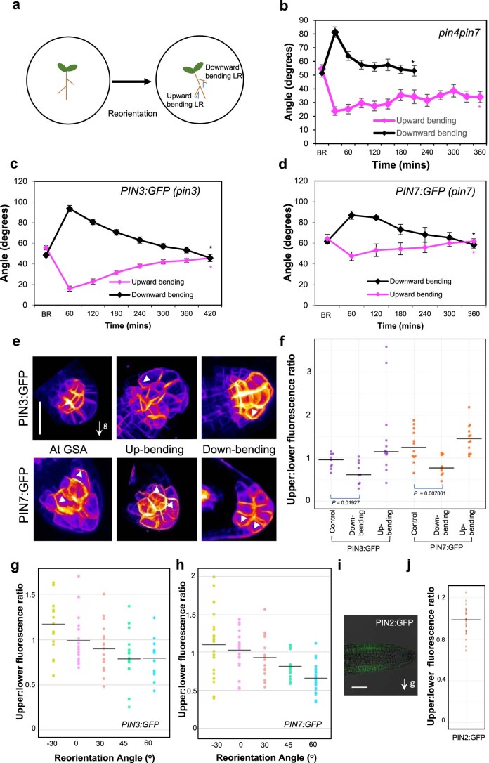
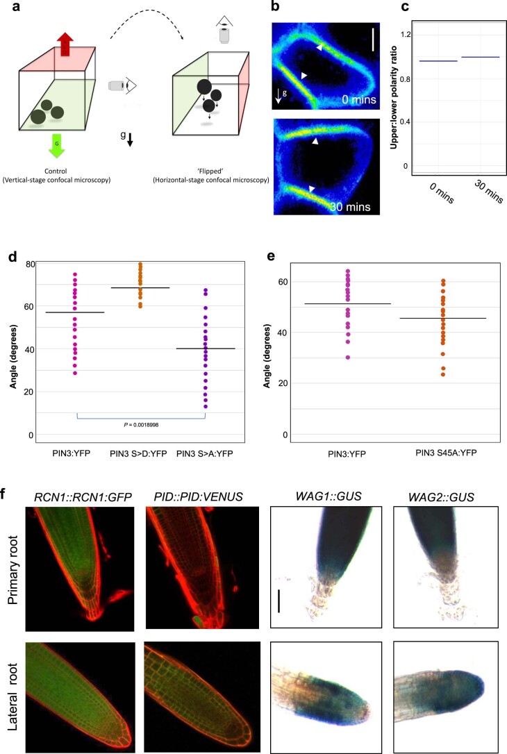
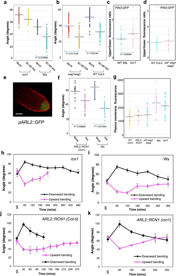
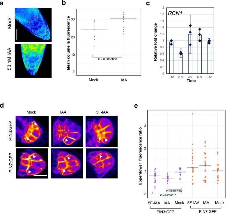
Lateral roots are typically maintained at non-vertical angles with respect to gravity. These gravitropic setpoint angles are intriguing because their maintenance requires that roots are able to effect growth response both with and against the gravity vector, a phenomenon previously attributed to gravitropism acting against an antigravitropic offset mechanism. Here we show how the components mediating gravitropism in the vertical primary root-PINs and phosphatases acting upon them-are reconfigured in their regulation such that lateral root growth at a range of angles can be maintained. We show that the ability of Arabidopsis lateral roots to bend both downward and upward requires the generation of auxin asymmetries and is driven by angle-dependent variation in downward gravitropic auxin flux acting against angle-independent upward, antigravitropic flux. Further, we demonstrate a symmetry in auxin distribution in lateral roots at gravitropic setpoint angle that can be traced back to a net, balanced polarization of PIN3 and PIN7 auxin transporters in the columella. These auxin fluxes are shifted by altering PIN protein phosphoregulation in the columella, either by introducing PIN3 phosphovariant versions or via manipulation of levels of the phosphatase subunit PP2A/RCN1. Finally, we show that auxin, in addition to driving lateral root directional growth, acts within the lateral root columella to induce more vertical growth by increasing RCN1 levels, causing a downward shift in PIN3 localization, thereby diminishing the magnitude of the upward, antigravitropic auxin flux.


Conflict of interest statement

The authors declare no competing interests.

Figures

Fig. 1
Fig. 1. Lateral root graviresponse is angle dependent and driven by auxin transport-dependent auxin asymmetry.
a,b, Mean GSA of mock- and NPA-treated stage III lateral roots growing at GSA and 24 h after reorientation by 30°. Treatment with 0.2 µM and 0.4 µM NPA inhibits lateral root reorientation in both upward (a) and downward (b) directions. n = 25–37 roots for each treatment from 3 biologically independent experiments. One-way ANOVA followed by post hoc Tukey’s HSD test revealed F(5) = 13.7890 for a and 25.8968 for b gave P = 1.102 × 10−16 for a and b. c, Change in length of stage III lateral roots during mock and NPA treatments. The growth rates of NPA-treated roots are not significantly different from mock-treated roots (P = 0.0838) n = 10 roots per treatment from 3 biologically independent experiments. d, Visualization of auxin fluxes using the auxin reporter DII-Venus in upward and downward reoriented lateral roots. Scale bar, 20 μm. e, Ratio of mean nuclear fluorescence between upper and lower epidermal cells of stage III lateral roots gravistimulated 30° above or below their GSA using the auxin reporter DII-Venus. Note: to aid understanding, the colloquial terms up- and down-bending are used as short descriptors of lateral roots undergoing negative (upward) and positive (downward) gravitropic response respectively. White arrowheads indicate increased nuclear fluorescent signal on the upper and lower sides of downward and upward bending lateral roots respectively. n = 10–15 roots analysed for each angle. Roots were reoriented from their GSA on the rotating stage of a vertical confocal microscope and imaged 60 min post reorientation. One-way ANOVA followed by post hoc Tukey’s HSD test revealed F(2) = 6.325 gave P value of 0.0039. f,g, Quantification of atrichoblast epidermal cell lengths at upper and lower sides of reorientated lateral roots. In upward-bending lateral roots, epidermal cells on the bottom half of the root are significantly longer than those on the upper side (f middle panel and g). In contrast, in downward-bending lateral roots, epidermal cells on the bottom of the root are significantly shorter in length than those on the upper side (f third panel and g). Scale bars, 50 μm. n = 20–35 for each group from 3 biologically independent experiments. One-way ANOVA followed by post hoc Tukey’s HSD test revealed F(5) = 13.7890 gave P value of 5.2996 × 10−11. h, Kinetics of gravitropism in lateral roots growing at oblique orientations. Curvature was measured in terms of stage rotation for roots maintained 30° from their original orientation by a feedback system following either an upward reorientation, resulting in positive gravitropism, or a downward reorientation, resulting in negative gravitropism (mean ± standard error of the mean, n = 18–22). i, Ratio of mean nuclear DII-Venus fluorescence between upper and lower epidermal cells of stage III lateral roots gravistimulated above or below their GSA by different angles. Roots were gravistimulated for 90 min before imaging. n = 8–10 roots for each angle of stimulation from 3 biologically independent experiments. Source data
Fig. 2
Fig. 2. PIN polarity distribution in lateral roots.
a,b, Comparison of PIN polarity in lateral roots at their GSA and primary roots reoriented by 45° roots in seedlings expressing PIN3:GFP (a) and PIN7:GFP (b). Fluorescence was measured on upper and lower membranes of outer columella cells as indicated in Extended Data Fig. 1c. PIN3 shows a slight polarity towards the lower cell membrane (a), while, in contrast, PIN7:GFP shows enhanced polarity towards the upper membrane (b) in lateral roots growing at their GSA. c, However, both PIN3:GFP (a) and PIN7:GFP (b) are predominantly polarized towards the lower plasma membrane in primary roots reoriented by ~45° for 30 min. Scale bar, 15 μm (a and b). n = 20–25 roots from 3 biologically independent experiments. Statistical analysis was carried out using a pairwise two-tailed t-test. di, Comparison of reorientation kinetics in lateral roots of 12-day-old WT Col-0 (d), pin3 pin7 (e), pin3pin4pin7 (f), pin3pin4 (g), pin3-3 (h) and pin7-2 (i) seedlings gravistimulated both above and below their GSA. BR represents GSA before reorientation. Average GSA of 10–12 upward- and downward -bending stage III lateral roots was quantified after reorientation until the roots were within 5° of their original GSA. Black asterisks indicate the timepoint at which angles were recovered for downward-bending roots, while magenta asterisks indicate the timepoint at which angles were recovered for upward-bending roots. pin3 and pin3pin4 lateral roots reorientate upwards significantly faster (g and h), while pin7 lateral roots reorientate downwards at a faster rate (i). WT Col-0 control lateral roots reorientate back to their GSA in both directions in approximately 6 h (d). In contrast, reorientation in both directions is delayed in the pin3 pin7 double mutant (e) and is virtually negligible in the pin3pin4pin7 triple mutant (f). n = 15–21 roots at all timepoints (di) from 3 biologically independent experiments. Bars in di represent standard error of the means.
Fig. 3
Fig. 3. PIN phosphorylation affects PIN polarity and redistribution kinetics in lateral root statocytes.
ad, PIN polarity distribution in columella cells of lateral roots rotated around their axis of growth by 90° (‘flip assays’; for a diagrammatic description of the experiment, see Extended Data Fig. 3a). In all panels, the former upper side of the columella cell is towards the top of the page. Post flip, phosphorylated PIN3 and PIN7 are retained on the upper plasma membrane for approximately 30 min longer than lower side unphosphorylated PINs. PIN3 (a and b) and PIN7 (c and d) polarity gradually becomes symmetrical on upper and lower sides of the plasma membrane 2 h after ‘flipping’ (d). Scale bar, 5 μm. n = 25–31 roots for each timepoint for b and d from 3 biologically independent experiments. One-way ANOVA followed by post hoc Tukey’s HSD test revealed F stat (3) = 3.8028 with a P value of 0.01027 for b and 5.8904 with a P value of 0.01456 for d. White arrowheads in a and c denote changes in PIN polarity over time. e,f, Quantification of PIN3 polarity in transgenic lines expressing non-phosphorylatable (S>A) or phosphomimic (S>D) variant of PIN3–YFP. Scale bar, 15 μm. n = 21 roots for each line from 3 biologically independent experiments. One-way ANOVA followed by post hoc Tukey’s HSD test revealed F stat (2) = 10.692 with a P value of 0.0001. g,h, PIN3:YFP polarity ratios after 30 min in horizontally flipped lateral roots in transgenic PIN3 phosphovariant lines. PIN3:YFP polarized to the upper side 30 min after flipping in WT PIN3 and PIN3 S>D: YFP phosphomimic lines, but not in the PIN3 S>A:YFP phosphodead line. Scale bar, 10 μm. n = 21 roots for each line from 3 biologically independent experiments. One-way ANOVA followed by post hoc Tukey’s HSD test revealed F stat (2) = 4.541 with a P value of 0.0498. i,j, Quantification of lateral root GSA phenotypes in rcn1 and wag1 wag2 mutants. rcn1 lateral roots have significantly less vertical lateral roots as compared with WT Ws seedlings (i). In contrast, wag1 wag2 seedlings have significantly more vertical lateral roots than WT Col-0 controls (j). n = 21 roots for each genotype from 3 biologically independent experiments (i and j). Statistical analysis was performed using two tailed t-tests (i and j).
Fig. 4
Fig. 4. Auxin regulates lateral root GSA through an RCN1-dependent PIN3 module.
a,b, Overexpression of RCN1 driven by the ARL2 promoter (ARL2::RCN1) in a WT Col-0 background results in a significantly more vertical lateral root GSA phenotype in contrast to Col-0 control (a) and restores the GSA of rcn1 lateral roots (b). n = 15–25 for each genotype from 3 biologically independent experiments for both a and b. Statistical analysis was performed using a two-tailed t-test for a and a one-way ANOVA with an F stat (2) = 19.3276 with P = 4.188 × 10−5. c,d, RCN1:GFP protein levels in 10-day-old lateral roots treated with 50 nM IAA for 4 h. Auxin treatment results in a significant increase in GFP signal in the columella cells of RCN1:GFP lateral roots. n = 15–21 roots per treatment from 3 biologically independent experiments. Statistical analysis was performed using a two tailed t-test (d). Red dashed lines represent columella area used for quantification (c). Scale bar, 20 μm (c). e,f, Visualisation (e) and quantification (f) of the effect of overexpression of RCN1 on PIN polarity in lateral root columella cells. RCN1 overexpression leads to a significant shift in PIN3:GFP polarity towards the lower side of the cell (d). In contrast, PIN7:GFP polarity is unaffected. n = 18–24 for each genotype from 3 biologically independent experiments. Statistical analysis was performed using a pairwise two-tailed t-test. Scale bar, 15 μm (e). g, The PP2A/RCN1 subunit is able to dephosphorylate the central hydrophilic loop of PIN3 in vitro. The experiment was repeated independently three times with similar results.
Fig. 5
Fig. 5. Model of auxin-dependent regulation of GSA.
a, Model of GSA control in the lateral root in which phosphorylated PIN3 and PIN7 mediate upward, antigravitropic auxin flux from columella cells, while unphosphorylated PIN3 and PIN7 mediate downward, gravitropic auxin transport. In addition to regulating cell elongation further back along the root, auxin also positively regulates levels of the PIN phosphatase subunit RCN1, thereby diminishing the magnitude of AGO. This causes the equilibrium between angle-dependent gravitropic- and angle-independent antigravitropic auxin flux to occur at a more vertical setpoint angle. b, Tropic response to displacement either above or below GSA is driven by angle-dependent changes in downward gravitropic auxin flux acting in tension with a more constant, angle-independent upward antigravitropic auxin flux. The thickness of red and green arrows signifies relative auxin flux.
Extended Data Fig. 1
Extended Data Fig. 1. Analysis of auxin signalling and PIN polarity in lateral root columella cells.
(a) Ratio of nuclear R2DII signal across upper and lower epidermal cells in lateral roots at GSA (control) and reorientated upwards and downwards. Images were taken 90 mins post reorientation Bars represent standard error of mean nuclear fluorescence. n = 12–15 roots for each orientation from 3 biologically independent experiments. One way ANOVA revealed F stat (2) = 22.25654 with a p value = 0.001346. (b) Statistical analysis using pairwise two-tailed T-tests revealed no significant difference in mean nuclear fluorescence of TIR1/AFB:Venus in atrichoblast cells on the upper and lower side of stage III lateral roots. n = 39–51 nuclei analysed for each transgenic line across 3 biologically independent experiments. (c) Outer membranes (white arrowheads) of upper and lower cells of the central columella used for quantification of PIN polarity in a single stack of a PIN3:GFP lateral root. Scale bar = 5 μm. (d) Quantification of PIN3/7::GFP fluorescence levels in plasma membranes of columella cells from stage III lateral and primary roots. n = 21–25 roots for each transgenic line quantified from 3 biologically independent experiments. Pairwise two tailed T-tests revealed no significant differences in membrane fluorescence levels.
Extended Data Fig. 2
Extended Data Fig. 2. Quantification of the kinetics of gravitropic response in lateral roots of single and multiple pin mutants.
(a) Schematic representation of seedling reorientation for analysis of reorientation kinetics in WT and pin mutants. Plates were reoriented by 30° angles and tip angles of roots placed above and below their GSA (denoted in blue) were quantified at specified time intervals. (b) Reorientation kinetics of 12-day-old lateral roots in the pin4pin7 double mutant. pin4pin7 lateral roots bend downwards rapidly, but show delayed upward bending presumably due to the loss of PIN7. (c, d) Reorientation kinetics of stage III lateral roots in the PIN3:GFP (C) and PIN7:GFP (D) transgenic lines. Lateral roots return to their original GSA in approximately 6 hours after reorientation in both directions. (B-D) n = 17–21 lateral roots at each time point from 3 biologically independent experiments (e, f) PIN polarity changes in 12-day old lateral roots reorientated in upward and downward directions. (e) PIN3:GFP and PIN7:GFP are predominantly dipolar (PIN3) and apolar (PIN7) in lateral roots at their GSA (right panels). In lateral roots reorientated below their GSA, both PIN3 and PIN7 polarise towards the upper side of columella cells (middle panels) whereas in roots displaced above their GSA, both PINs polarise towards the lower side of columella cells (left panels). White arrowheads indicate polar localisation of PIN:GFP signal. Scale bar = 5 μm. n = 12–15 roots at each reorientation from 3 biologically independent experiments. One way ANOVA revealed F stat (2) = 4.5096 with a P value = 0.0201 for PIN3:GFP and F stat (2) = 10.4650 with a P value = 0.0004 for PIN7:GFP. (g, h) Quantification of upper/ lower mean PIN3/7:GFP signal across external columella cell membranes in lateral roots at GSA and reorientated upwards and downwards at defined angles. Negative angles denote reorientation below GSA. Bars represent standard error of the means. (i) PIN2:GFP signal is not differentially expressed across upper and lower sides of lateral roots (right panel). Scale bar = 20 μm. (j) Quantification of PIN2:GFP upper/lower epidermal signal in trichoblasts across upper and lower epidermal cell files.
Extended Data Fig. 3
Extended Data Fig. 3. Phosphorylation influences membrane retention kinetics and polarity of PIN proteins in lateral root columella cells.
a) Schematic representation of the ‘flip’ assay designed to study PIN membrane retention kinetics using vertical stage microscopy. (b, c) The PIP1;4:YFP (Wave11_Y) plasma membrane marker remains apolar in columella cells at their GSA (0 mins) or 30 mins after ‘flipping’. Scale bar = 5 μm. White arrowheads denote fluorescent signal at the plasma membrane (b). (c) Ratio of PIP1;4:YFP plasma membrane polarity in lateral root columella cells at GSA and 30 mins post ‘flipping’. n = 12–15 roots for each time point from 3 biologically independent experiments. (d) Quantification of GSA phenotypes in lateral roots of 12-day-old PIN3:YFP S > A and S > D phosphovariant lines. (e) Quantification of lateral root GSA in the 12-day-old seedlings of PIN3:YFP D6PK phosphovariant line (PIN3:S45A:YFP). n = 15–21 roots for each genotype from 3 biologically independent experiments for (d) and (e). One way ANOVA with an F stat (2) = 7.7295 revealed a p value = 0.0026 for (d) while, a two tailed T-test revealed no significant differences were observed as compared to the PIN3:YFP control (e). (f) Expression of PP2AA/RCN1 phosphatase subunit and the PID/WAG kinase family in primary and lateral roots. RCN1::RCN1:GFP is expressed in both, the primary and lateral root columella cells (left upper and lower panels). In contrast PID::PID:VENUS and WAG1::GUS are not expressed in the primary or lateral root columella (Centre upper and lower panels). WAG2::GUS is absent from the primary root columella, but strongly expressed in the lateral root columella. Scale bar = 30 μm. The experiment was repeated independently three times with similar results.
Extended Data Fig. 4
Extended Data Fig. 4. PIN3 dephosphorylation via RCN1 regulates lateral root GSA.
(a,b) Effect of 50 nM auxin treatment on rcn1 (a and wag1wag2 (b) mutant lateral roots. rcn1 lateral roots are unaffected by auxin treatment. wag1wag2 lateral roots adopt a more vertical orientation upon auxin treatment. n = 12–15 roots per genotype per treatment for (A) and (B). One way ANOVA revealed an F stat (3) of 18.0109 and a p value = 5.09X10⁻⁶ for (a) and an F stat (3) = 10.1844 and a p value = 0.0002 for (b). (c, d) Upper/lower membrane ratios of PIN3:GFP in the columella cells of the rcn1 and pid+wag1wag2 mutants. PIN3:GFP polarity is shifted to the upper columella membrane in the rcn1 background but remains unaffected in the pid+wag1wag2 background. n = 24–27 roots for each genotype from 3 biologically independent experiments for (c) and (d). Data was statistically analysed using a two tailed T-test. (e) Expression of ARL2::GFP in lateral root columella cells. Scale bar = 50 µm. (f) Auxin treatment (50 nM IAA) shifts the lateral root GSA of ARL2::RCN1 rcn1 lateral roots to a significantly steeper orientation. n = 12–15 roots for each genotype per treatment from 3 biologically independent experiments. One way ANOVA revealed an F stat(3) value = 15.8911 with a p value = 3.67X10⁻⁷. (g) Quantification of PIN3:GFP fluorescence in the columella cell plasma membranes in WT and mutant backgrounds. Fluorescence levels did not significantly differ between mutants and their WT controls. n = 22–30 per genotype from 3 biologically independent experiments. (h) Stage III lateral roots in the rcn1 mutant background reorientate upwards to their original GSA in approximately 4 hours, while downward reorientation is delayed. (i) Stage III lateral roots of WT Ws plants reorientate both upwards and downwards in 6 hours. (j) In contrast, overexpression of RCN1 in the columella in the WT Col-0 background leads to rapid downward reorientation of stage III lateral roots. (k) However, expression of RCN1 in the rcn1 mutant background restores reorientation kinetics of stage III lateral roots to a similar pattern as Ws. Data represent average values from 3 independent experiments. Bars represent standard errors of the means (h-k) with 6-8 roots reorienting in each direction per experiment per time point.
Extended Data Fig. 5
Extended Data Fig. 5. Effect of auxin treatment on RCN1 expression and protein stability.
a,b) Effect of auxin treatment for 4 hours on RCN1::RCN1:GFP (PP2AA::PP2AA:GFP translational reporter line) primary root columella cells. Auxin treatment leads to a significant increase in RCN1:GFP signal levels. n = 15–21 roots from 3 biologically independent experiments. Statistical analysis was performed using a two tailed T-test. Scale bar = 20 µm. Red dashed lines represent area of columella signal quantification in (a). (c) Effect of 50 nM IAA on RCN1 transcript levels in lateral root columella cells. No significant increase in RCN1 levels occurred over an 8 hour time course. Data represent averages from 3 independent experiments with 7-8 root tips harvested for each time point per experiment. Bars represent standard error of the means. (d,e) Treatment with 50 nM IAA or 5F-IAA results in a shift in PIN3:GFP polarity towards the lower side of the columella cell, but has no effect on PIN7:GFP polarity. n = 12–15 roots per treatment from 3 biologically independent experiments. One way ANOVA revealed an F stat(5) value = 4.9116 with a P value = 0.0130.

Comment in

References

    1. Digby RD, Firn J. The gravitropic set-point angle (GSA): the identification of an important developmentally controlled variable governing plant architecture. Plant Cell Environ. 1995;18:1434–1440. doi: 10.1111/j.1365-3040.1995.tb00205.x. - DOI - PubMed
    1. Nemec B. Ueber die Art der Wahrnehmung des Schwerkraftreizes bei den Pflanzen. Ber. Dtsch Bot. Ges. 1900;18:241–245. doi: 10.1111/j.1438-8677.1900.tb04905.x. - DOI
    1. Haberlandt G. Ueber die Perzeption des geotropischen Reizes. Ber. Dtsch Bot. Ges. 1900;18:261–272. doi: 10.1111/j.1438-8677.1900.tb04908.x. - DOI
    1. Sack FD, Kiss JZ. Rootcap structure in wild-type and in a starchless mutant of Arabidopsis. Am. J. Bot. 1989;76:454–464. doi: 10.1002/j.1537-2197.1989.tb11334.x. - DOI - PubMed
    1. Cholodny N. Wuchshormone und Tropismem bei den Pflanzen. Biol. Zentralbl. 1927;47:604–626.

Publication types

Substances