Review
. 2023 Sep 4;21(1):227.
doi: 10.1186/s12964-023-01247-5.

Regulation of anoikis by extrinsic death receptor pathways

Affiliations
Review

Regulation of anoikis by extrinsic death receptor pathways

Ying-Hao Han et al. Cell Commun Signal. .

Abstract

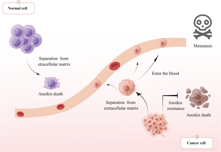
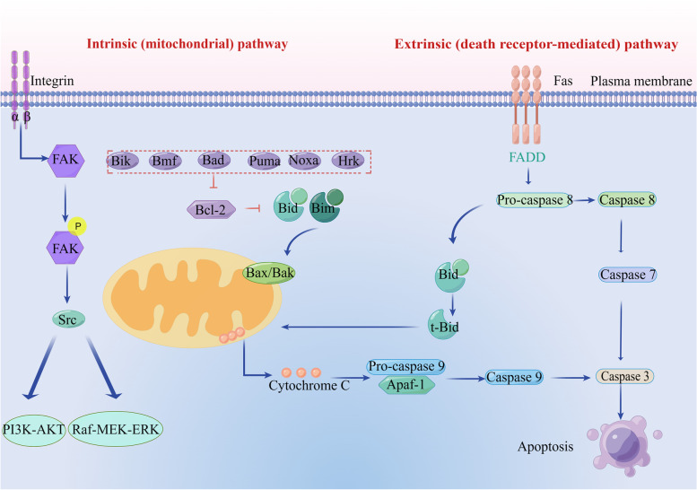
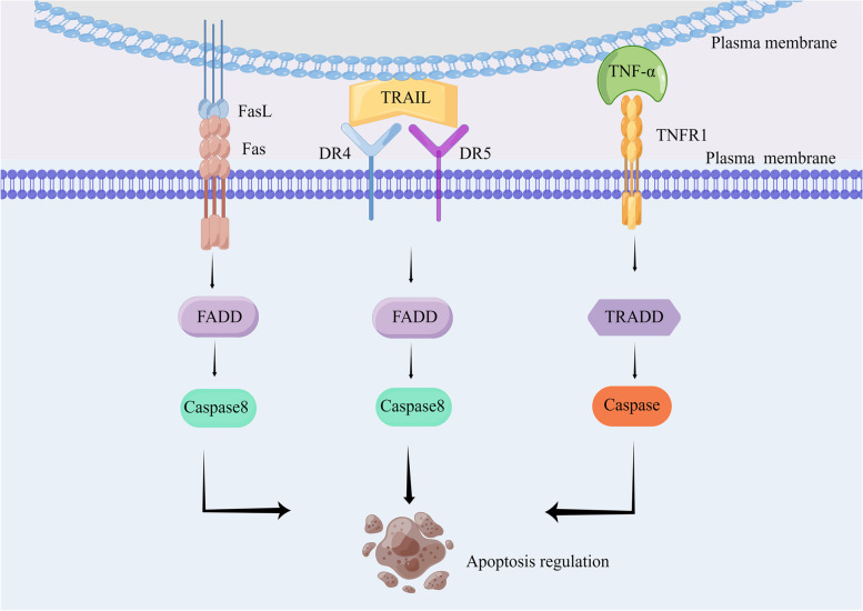
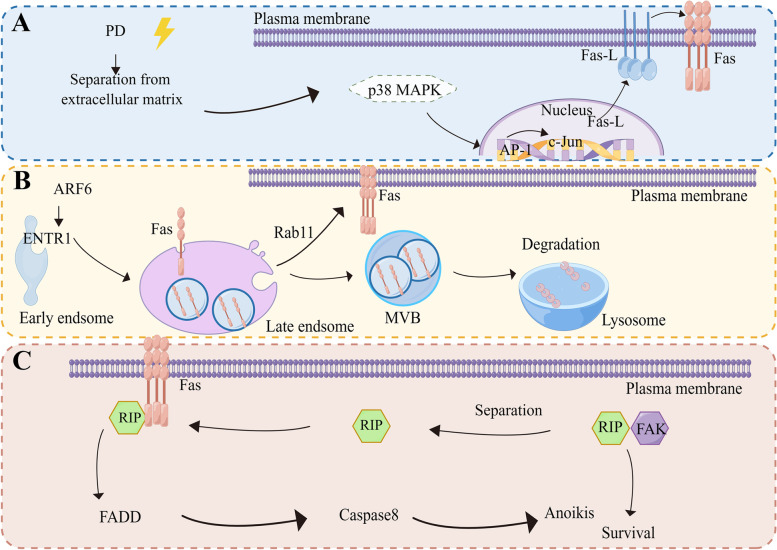
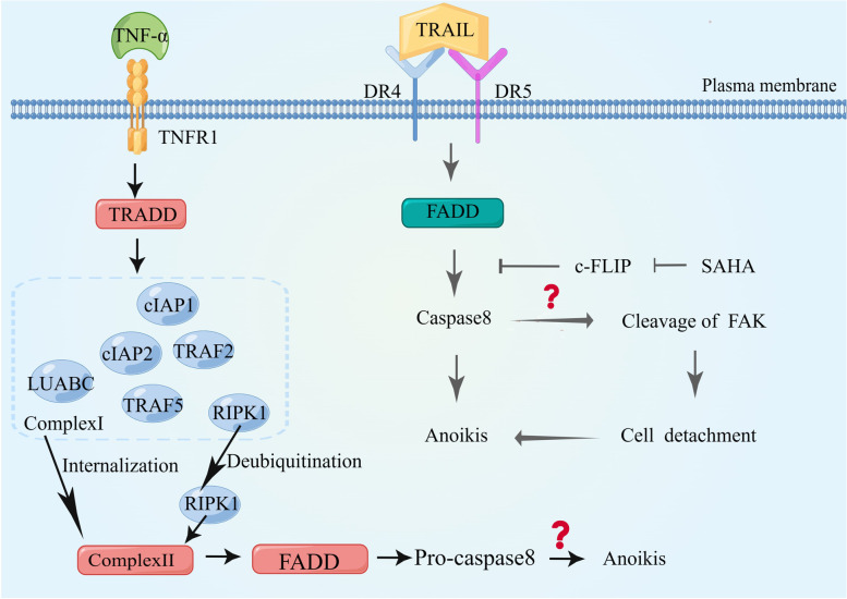
Metastatic cancer cells can develop anoikis resistance in the absence of substrate attachment and survive to fight tumors. Anoikis is mediated by endogenous mitochondria-dependent and exogenous death receptor pathways, and studies have shown that caspase-8-dependent external pathways appear to be more important than the activity of the intrinsic pathways. This paper reviews the regulation of anoikis by external pathways mediated by death receptors. Different death receptors bind to different ligands to activate downstream caspases. The possible mechanisms of Fas-associated death domain (FADD) recruitment by Fas and TNF receptor 1 associated-death domain (TRADD) recruitment by tumor necrosis factor receptor 1 (TNFR1), and DR4- and DR5-associated FADD to induce downstream caspase activation and regulate anoikis were reviewed. This review highlights the possible mechanism of the death receptor pathway mediation of anoikis and provides new insights and research directions for studying tumor metastasis mechanisms. Video Abstract.

Keywords: Aanoikis; Cancer metastasis; DR4/DR5; Death receptor pathway; Fas; TNFR1/TNFR2.


Conflict of interest statement

The authors declare no competing interests.

Figures

Fig. 1
Fig. 1
Cell fate of normal cells and metastatic tumor cells after losing adhesion to the extracellular matrix. When normal cells lose their adhesion to the extracellular matrix, apoptosis occurs because the body prevents the wrong connection of cells. However, after losing adhesion to the extracellular matrix due to its metastasis characteristics, tumor cells develop resistance, survive without adhesion to the extracellular matrix, and can flow to other tissues and organs through blood and lymph for adhesion and metastasis
Fig. 2
Fig. 2
Pathway diagram of internal and external mechanisms of anoikis. Lack of ECM exposure or inappropriate ECM exposure fails to activate pro-survival signaling, leading to a reduction in the anti-apoptotic pathway and, thus, activation of extrinsic death receptors and intrinsic integrin-mediated mitochondria-dependent anoikis. The death receptor pathway triggers two different extrinsic pathways in different cell types: type 1 leads to direct proteolysis of the target dependent on caspase-7 activation; type 2, which aggregates in the intrinsic pathway through a truncated form of Bid (tBid), promotes the release of mitochondrial cytochrome c and assembly of apoptotic corpuscles. These events induce proteolysis of caspase-specific targets, thereby promoting anoikis
Fig. 3
Fig. 3
Construction of anoikis model to construct an anoikis model, different approaches can be used, including the use of specialized cell culture dishes and various drugs. Common drugs that induce apoptosis include H2O2, tunicamycin, thapsigargin, staurosporine, and TNF-α. These drugs can induce anoikis through different mechanisms, such as oxidative stress, endoplasmic reticulum stress, and protein aggregation. Therefore, the selection of these drugs should be based on specific experimental conditions and research purposes
Fig. 4
Fig. 4
Binding of different death receptors to ligands and downstream signal transduction. Fas and FasL combine to activate downstream FADD and cause caspase activation. TNFR binds to TNFR1 to recruit TRADD, and post-translational modification of RIPK1 determines cell apoptosis. DR4 and DR5 are involved in an external pathway mediating caspase-8 activation
Fig. 5
Fig. 5
Three possible mechanisms of Fas-mediated anoikis. Elevated Fas expression levels result in the formation of downstream FADD and activation of caspase. A Firstly, the increase in the transcription level of Fas ligand FasL leads to the increase in the transcription level of receptor Fas and induces the formation of FADD. B Secondly, the increased expression level of Fas may be due to the decreased degradation of lysosomes through vesicle sorting through endocytosis, which leads to the increased accumulation of Fas in endosomes and the increased location of Fas to the plasma membrane surface dependent on Rab11-mediated recirculation and activation of downstream FADD. C The third is that RIP shuttles between Fas and FAK to regulate anoikis. RIP can form complexes with Fas or FAK to activate different signaling pathways under different conditions
Fig. 6
Fig. 6
Possible mechanisms of anoikis mediated by TNFR1 and DR4/DR5. TNF-α binding to TNFR1 leads to the formation of different signaling complexes that define different cell fates. TNF-α binding to TNFR1 recruits TNF receptor-associated proteins and the death domain (TRADD). TRADD forms signal complex I with TRAF2, TRAF5, RIPK1, LUBAC, cIAP1, and cIAP2. Complex I internalizes and transforms into the death-inducing complex, Complex II, with additional recruitment of FADD and pro-caspase8. Many outcomes of TNFR1 activation depend on post-translational modification of Complex I member RIPK1; deubiquitination of RIPK1 facilitates its release from Complex I and binding to Complex II to induce apoptosis. DR5 and DR4 can participate in anoikis, but the specific mechanism remains to be clarified

References

    1. Simpson CD, Anyiwe K, Schimmer AD. Anoikis resistance and tumor metastasis. Cancer Lett. 2008;272:177–185. doi: 10.1016/j.canlet.2008.05.029. - DOI - PubMed
    1. Su H, Si X-Y, Tang W-R, Luo Y. The regulation of anoikis in tumor invasion and metastasis. Yi Chuan = Hereditas. 2013;35:10–16. doi: 10.3724/SP.J.1005.2013.00010. - DOI - PubMed
    1. Kakavandi E, Shahbahrami R, Goudarzi H, Eslami G, Faghihloo E. Anoikis resistance and oncoviruses. J Cell Biochem. 2018;119:2484–2491. doi: 10.1002/jcb.26363. - DOI - PubMed
    1. Raeisi M, Zehtabi M, Velaei K, Fayyazpour P, Aghaei N, Mehdizadeh A. Anoikis in cancer: The role of lipid signaling. Cell Biol Int. 2022;46:1717–1728. doi: 10.1002/cbin.11896. - DOI - PubMed
    1. Zhu Z, Fang C, Xu H, Yuan L, Du Y, Ni Y, Xu Y, Shao A, Zhang A, Lou M. Anoikis resistance in diffuse glioma: The potential therapeutic targets in the future. Front Oncol. 2022;12:976557. doi: 10.3389/fonc.2022.976557. - DOI - PMC - PubMed