. 2023 Sep 4;14(1):236.
doi: 10.1186/s13287-023-03478-2.

Low-glucose culture environment can enhance the wound healing capability of diabetic adipose-derived stem cells

Affiliations

Low-glucose culture environment can enhance the wound healing capability of diabetic adipose-derived stem cells

Chun-Wei Li et al. Stem Cell Res Ther. .

Abstract

Background: Application of autologous adipose-derived stem cells (ASC) for diabetic chronic wounds has become an emerging treatment option. However, ASCs from diabetic individuals showed impaired cell function and suboptimal wound healing effects. We proposed that adopting a low-glucose level in the culture medium for diabetic ASCs may restore their pro-healing capabilities.

Methods: ASCs from diabetic humans and mice were retrieved and cultured in high-glucose (HG, 4.5 g/L) or low-glucose (LG, 1.0 g/L) conditions. Cell characteristics and functions were investigated in vitro. Moreover, we applied diabetic murine ASCs cultured in HG or LG condition to a wound healing model in diabetic mice to compare their healing capabilities in vivo.

Results: Human ASCs exhibited decreased cell proliferation and migration with enhanced senescence when cultured in HG condition in vitro. Similar findings were noted in ASCs derived from diabetic mice. The inferior cellular functions could be partially recovered when they were cultured in LG condition. In the animal study, wounds healed faster when treated with HG- or LG-cultured diabetic ASCs relative to the control group. Moreover, higher collagen density, more angiogenesis and cellular retention of applied ASCs were found in wound tissues treated with diabetic ASCs cultured in LG condition.

Conclusions: In line with the literature, our study showed that a diabetic milieu exerts an adverse effect on ASCs. Adopting LG culture condition is a simple and effective approach to enhance the wound healing capabilities of diabetic ASCs, which is valuable for the clinical application of autologous ASCs from diabetic patients.

Keywords: Adipose-derived stem cell; Diabetes; Stem cell culture; Stem cell transplantation; Tissue regeneration.


Conflict of interest statement

The authors declare that they have no competing interests.

Figures
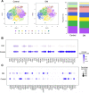
Fig. 1
Fig. 1
Single-cell RNA sequencing analysis of hASCs from non-diabetic (control) and diabetic (DM) patients. A Cell population distribution by t-SNE projections showed a similar pattern in control and DM individuals (left and middle, n = 8812 and 8689, respectively). Bar charts of 11 clusters identified from control and diabetic ASCs showed slight differences in the proportion of clusters (right). B Dot plot showed decreased expression of the several ECM organizing-related genes in hASCs from DM patients. C Dot plot showed variation in the relative expression of angiogenesis-related genes in hASCs from both groups. The sizes of the dots in (B) and (C) corresponded to the percentage of cells expressing the genes in each cluster, and the color scale represented the average expression level. hASC human adipose-derived stem cell, ECM extracellular matrix, t-SNE t-distributed stochastic neighbor embedding
Fig. 2
Fig. 2
Characteristics of control hASCs cultured in high- or low-glucose conditions. A Cumulative population doubling of hASCs revealed a lower proliferative activity when cultured in HG condition, and the cell growth could be enhanced after shifting to LG condition at day 35 (arrow). B Proliferative activity estimated by BrdU incorporation showed faster growth of hASCs in the 35H14L and 49L groups. C Cytotoxicity of hASCs in the serum-free condition determined by the LDH efflux assay. D SA-β-gal staining of hASCs and their quantification in five randomly selected power fields. Scale bar = 100 μm. E Cell migration of hASCs evaluated by in vitro scratch assay at different time points. Scale bar = 100 μm. *p < 0.05, **p < 0.01 relative to the 49H group; #p < 0.05, ##p < 0.01 between the indicated groups. BrdU 5-bromo-2′-deoxyuridine, hASC human adipose-derived stem cell, HG high glucose, LG low glucose, hpf high-power field, SA-β-gal senescence-associated β-galactosidase, 49H 49 days of high-glucose condition, 49L 49 days of low-glucose condition, 35H14L 35 days of high-glucose condition followed by 14 days of low-glucose condition
Fig. 3
Fig. 3
Phenotypic characterization of murine ASCs from db/db mice in high-glucose (dbHG) or low-glucose (dbLG) conditions. A The expression level of ASC surface markers is shown as the proportion of positively stained cells relative to the isotype control. Both groups of ASCs were negative for the hematopoietic markers CD31 and CD34 but positive for the mesenchymal stem cell-related markers CD90. B Murine ASCs were cultured in adipogenic or osteogenic induction medium, and cells were stained with Oil Red O for the detection of adipogenesis and Alizarin Red for the detection of osteogenesis. Scale bar = 200 μm. C Cumulative population doubling curve showing faster proliferation of the dbLG group. D Quantification of SA-β-gal-positive cells in five randomly selected power fields revealed more senescent cells in the dbHG group. E After cultured in LG and HG conditions, murine ASCs were examined for ROS production. Significantly more ROS production was noted in the dbHG condition. *p < 0.05, **p < 0.01 relative to dbHG. ASC adipose-derived stem cell, SA-β-gal senescence-associated β-galactosidase, ROS reactive oxygen species
Fig. 4
Fig. 4
Expression of angiogenic growth factors in dbASCs under high-glucose (dbHG) and low-glucose (dbLG) conditions. A Evaluation of angiogenesis‑related gene expression by quantitative RT-PCR, including FGF2, VEGF, and HGF. Values are normalized by GAPDH expression level and plotted as relative quantity to the dbHG group. *p < 0.05 relative to dbHG. B ELISA showed significantly higher concentration of HGF in CM-dbLG. *p < 0.05 relative to CM-dbHG. C BrdU assay demonstrated enhanced proliferative activity of HUVECs incubated in CM-dbLG. **p < 0.01 relative to CM-dbHG. D In vitro tube formation of HUVECs. HUVECs cultured in endothelial growth medium 2 (EGM2) served as positive controls, and those in endothelial basal medium 2 (EBM2) were negative controls. Scale bar = 500 μm. Branching nodes, junctions and segments per power field were compared among different groups except the positive control. *p < 0.05, **p < 0.01 relative to control; #p < 0.05, ##p < 0.01 between the indicated groups. E In vitro scratch assay of human dermal fibroblasts. Red dotted lines represent the initial borders of the cell-free zone. Scale bar = 200 μm. Quantification of the wound closure percentage of fibroblasts was shown. *p < 0.05, **p < 0.01 relative to CM-dbHG. dbASC murine ASCs from db/db mice, CM-dbHG conditioned medium from dbASCs cultured in HG condition, CM-dbLG conditioned medium from dbASCs cultured in LG condition, HUVEC human umbilical vein endothelial cell
Fig. 5
Fig. 5
Application of murine ASCs from db/db mice in high-glucose (dbHG) or low-glucose (dbLG) conditions in a diabetic wound healing model. A Gross pictures of wounds applied with PBS, dbHG, or dbLG. B Relative wound area curves of the dbHG and dbLG groups compared with the contralateral wounds that received PBS treatment. *p < 0.05, **p < 0.01 relative to the PBS group. dbASC murine ASCs from db/db mice, dbHG dbASCs cultured in HG condition, dbLG dbASCs cultured in LG condition
Fig. 6
Fig. 6
Histology of the healed wound tissue. A Representative images of H&E-stained wound sections at day 21. Red arrows indicated the wound margin. Scale bar = 500 µm. B Representative images of Masson’s trichrome-stained wound sections at day 21. The differences in the dermal matrix organization were examined under higher magnification, and wounds treated with dbLG exhibited significantly higher collagen density relative to those received PBS or dbHG. Scale bar = 500 µm; higher magnification scale bar = 100 µm. *p < 0.05 relative to PBS, #p < 0.05 between the indicated groups. dbASC murine ASCs from db/db mice, dbHG dbASCs cultured in HG condition, dbLG dbASCs cultured in LG condition, H&E hematoxylin and eosin
Fig. 7
Fig. 7
Immunohistochemistry and dbASC retention in the wound tissue. A Representative images of the CD31 immunohistochemical sections of wound explants at day 5 and day 21. Red arrows indicate CD31-expressing cells around small vessels. Quantitative analysis of the CD31-expressing cells in high-power field images was shown. Scale bar = 50 µm. *p < 0.05 relative to PBS. B Representative images of wound sections stained with anti-CD68 and anti-α-SMA at day 21. Scale bar = 50 µm. Quantitative analysis of the immunohistochemical signals of CD68 and α-SMA showed no differences among different groups. C Residual dbASCs in the wound tissues at day 5 were visualized by Qtracker labeling. Scale bar = 100 µm. Significantly more retaining dbASCs were found in the wound tissue treated with dbLG. **p < 0.01 relative to the dbHG group. dbASC murine ASCs from db/db mice, dbHG dbASCs cultured in HG condition, dbLG dbASCs cultured in LG condition

Similar articles

Cited by

References

    1. Ahmad E, Lim S, Lamptey R, et al. Type 2 diabetes. Lancet. 2022;400(10365):1803–1820. doi: 10.1016/S0140-6736(22)01655-5. - DOI - PubMed
    1. Armstrong DG, Boulton AJM, Bus SA. Diabetic foot ulcers and their recurrence. N Engl J Med. 2017;376(24):2367–2375. doi: 10.1056/NEJMra1615439. - DOI - PubMed
    1. Spampinato SF, Caruso GI, De Pasquale R, et al. The treatment of impaired wound healing in diabetes: looking among old drugs. Pharmaceuticals (Basel) 2020;13(4):60. doi: 10.3390/ph13040060. - DOI - PMC - PubMed
    1. Bertozzi N, Simonacci F, Grieco MP, et al. The biological and clinical basis for the use of adipose-derived stem cells in the field of wound healing. Ann Med Surg (Lond) 2017;20:41–48. doi: 10.1016/j.amsu.2017.06.058. - DOI - PMC - PubMed
    1. Gentile P, Sterodimas A, Pizzicannella J, et al. Systematic review: allogenic use of stromal vascular fraction (SVF) and decellularized extracellular matrices (ECM) as advanced therapy medicinal products (ATMP) in tissue regeneration. Int J Mol Sci. 2020;21(14):4982. doi: 10.3390/ijms21144982. - DOI - PMC - PubMed

Publication types