. 2023 Oct;10(30):e2303283.
doi: 10.1002/advs.202303283. Epub 2023 Sep 5.

NKRF in Cardiac Fibroblasts Protects against Cardiac Remodeling Post-Myocardial Infarction via Human Antigen R

Affiliations

NKRF in Cardiac Fibroblasts Protects against Cardiac Remodeling Post-Myocardial Infarction via Human Antigen R

Chenghu Guo et al. Adv Sci (Weinh). 2023 Oct.

Abstract

Myocardial infarction (MI) remains the leading cause of death worldwide. Cardiac fibroblasts (CFs) are abundant in the heart and are responsible for cardiac repair post-MI. NF-κB-repressing factor (NKRF) plays a significant role in the transcriptional inhibition of various specific genes. However, the NKRF action mechanism in CFs remains unclear in cardiac repair post-MI. This study investigates the NKRF mechanism in cardiac remodeling and dysfunction post-MI by establishing a CF-specific NKRF-knockout (NKRF-CKO) mouse model. NKRF expression is downregulated in CFs in response to pathological cardiac remodeling in vivo and TNF-α in vitro. NKRF-CKO mice demonstrate worse cardiac function and survival and increased infarct size, heart weight, and MMP2 and MMP9 expression post-MI compared with littermates. NKRF inhibits CF migration and invasion in vitro by downregulating MMP2 and MMP9 expression. Mechanistically, NKRF inhibits human antigen R (HuR) transcription by binding to the classical negative regulatory element within the HuR promoter via an NF-κB-dependent mechanism. This decreases HuR-targeted Mmp2 and Mmp9 mRNA stability. This study suggests that NKRF is a therapeutic target for pathological cardiac remodeling.

Keywords: NF-κB-repressing factor; cardiac fibroblasts; human antigen R; myocardial infarction; transcriptional and post-transcriptional regulation.


Conflict of interest statement

The authors declare no conflict of interest.

Figures
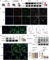
Figure 1
Figure 1
Downregulation of NKRF expression in cardiac fibroblasts (CFs) of the myocardial infarction (MI) border zone. A–C) Male C57BL/6J mice (aged 8 weeks) were subjected to MI by ligation of the left anterior descending coronary artery and euthanized at 28 days post‐MI. A) MI in mice. B) Western blotting and quantification of NKRF from the protein extracted within the MI border region in mice (n = 6). C) Immunofluorescence co‐staining of NKRF (red) and FSP1 (green) in the border zone of MI in mice (scale bar = 20 µm). D) Immunofluorescence staining shows the location of NKRF (green) in primary CFs (scale bar = 100 µm). E) Serum TNF‐α, IL‐1β, and IL‐6 levels in patients with ST‐segment elevation MI and normal healthy individuals. F–H) Primary CFs were treated with TNF‐α (10 ng mL−1) at the indicated time points. F) Western blotting and quantification of NKRF (n = 5). G) Immunoblotting and quantification of NKRF from isolated nuclear and cytoplasmic subcellular fractions of CFs (n = 6). H) Immunofluorescence staining of NKRF (green) in CFs treated with TNF‐α for 24 h (scale bar = 100 µm). Data are expressed as the mean ± SEM. NS, non‐significant, *p<0.05, and ****p<0.0001 (unpaired two‐tailed Student's t‐test). LAD, left anterior descending coronary artery; NKRF, NF‐κB‐repressing factor; FSP1, fibroblast‐specific protein 1.
Figure 2
Figure 2
NKRF protects against cardiac remodeling and dysfunction post‐MI. A) Schematic diagram depicting the time course of MI‐induced cardiac remodeling and dysfunction in NKRFF/F and NKRF‐CKO mice. B) Echocardiography and measured LVIDd, LVIDs, EF%, and FS% in NKRFF/F and NKRF‐CKO mice (n = 6). C) Cardiac magnetic resonance (CMR) imaging at the left ventricular end‐diastolic phase (LVED) and end‐systolic phase (LVES) in NKRFF/F and NKRF‐CKO mice. D) Masson's Trichrome (MT) and Picrosirius Red (PSR) staining from transverse cross‐sections of hearts obtained from NKRFF/F and NKRF‐CKO mice, along with infarct size quantification (scale bar = 1000 µm, n = 7). E) Ratio of heart weight to body weight (HW/BW) of mice post‐MI 28 days (n = 10). F) Immunoblot and quantification of MMP2, MMP9, Collagen I, and Collagen III from the MI border region protein (n = 6). G) Kaplan–Meier survival analysis of NKRF‐CKO (n = 40) and NKRFF/F mice (n = 20) after MI or sham operation. H) Deceased mice post‐MI; a. illustrates the hemothorax caused by cardiac rupture and b. shows the development of pulmonary edema. Data are expressed as the mean ± SEM. NS, non‐significant, *p<0.05, **p<0.01, ***p<0.001, and ****p<0.0001 by two‐way analysis of variance (ANOVA) with a Bonferroni multiple comparison test B,D,E,F) and log‐rank test G). NKRFF/F and NKRF‐CKO, NKRFflox/flox and NKRFflox/flox:CreS100a4 mice; LAD, left anterior descending coronary artery; MI, myocardial infarction; LVIDd, left ventricular internal diastolic dimension; LVIDs, left ventricular internal systolic dimension; EF, left ventricular ejection fraction; FS, fractional shortening; MMP2, matrix metalloproteinase 2; MMP9, matrix metalloproteinase 9.
Figure 3
Figure 3
NKRF inhibits CF migration and invasion by downregulating MMP2 and MMP9 expression. A) Transwell invasion assay. B–H) Primary CFs transfected with Ad‐Nkrf or Ad‐Vector for 24 h were treated with TNF‐α (10 ng mL−1) for another 24 h. B) Transwell invasion staining and relative migration rate calculation (scale bar = 50 µm, n = 7). C) Wound healing assays and migration rate quantification at the indicated time points (scale bar = 200 µm, n = 7). D–F) Relative mRNA expression of Nkrf, Mmp2, and Mmp9, respectively (n = 5). G) Western blotting and quantification of NKRF (n = 8), MMP2 (n = 7), and MMP9 (n = 6). H) Gelatin zymography and quantitative analysis of MMP2 and MMP9 activities in cultured supernatants (n = 4). Data are the mean ± SEM. *p<0.05, **p<0.01, ***p<0.001, and ****p<0.0001 (two‐way ANOVA with Bonferroni multiple comparisons test). Ad‐Nkrf, NKRF adenovirus.
Figure 4
Figure 4
NKRF inhibits the stability of Mmp2 and Mmp9 mRNA by inhibiting HuR expression. A) NKRF inhibits mRNA stability of Mmp2 (n = 6) and Mmp9 (n = 6). Significant differences were observed at 12 h. (B,C) NKRF inhibits TNF‐α‐induced HuR expression in CFs at the mRNA (B, n = 5) and protein (C, n = 7) levels. (D,E) NKRF knockdown enhances TNF‐α‐induced HuR expression in CFs at the mRNA (D, n = 5) and protein levels (E, n = 4). F) Western blotting and quantification of HuR protein extracted from the MI border region in NKRFF/F mice and NKRF‐CKO mice (n = 6). (G,H) Agarose gel electrophoresis and RT‐PCR results of Mmp2 (G, n = 4) and Mmp9 (H, n = 4) mRNA enriched by the HuR antibody in RIP experiments. (I,J) HuR rescues the NKRF‐inhibited mRNA stability of Mmp2 (I, n = 4) and Mmp9 (J, n = 4). Significant differences were observed at 12 h. Data are the mean ± SEM. NS, non‐significant, *p<0.05, **p<0.01, ***p<0.001, and ****p<0.0001 by unpaired two‐tailed Student's t‐test (A), two‐way ANOVA with Bonferroni multiple comparisons test B–F), and one‐way ANOVA with Bonferroni multiple comparisons test G–J). Ad‐Nkrf, NKRF‐overexpressing adenovirus; Ad‐HuR, HuR‐overexpressing adenovirus; SiR‐Nkrf, Nkrf siRNA (small interfering RNA); NKRFF/F, NKRFflox/flox mice; NKRF‐CKO, NKRFflox/flox:CreS100a4 mice; MI, myocardial infarction; Anti‐HuR, HuR antibody.
Figure 5
Figure 5
NKRF inhibits HuR transcription by binding to the HuR promoter via an NF‐κB dependent mechanism. A) Negative regulatory element (NRE) in the HuR promoter region. B,C) Agarose gel electrophoresis B) and RT‐PCR results (C, n = 4) using NRE in the HuR promoter region enriched with NKRF antibody as a template in chromatin immunoprecipitation (ChIP) experiments. D) Firefly luciferase expression plasmid construction with the wild‐type HuR promoter (WT, pGL3‐WT‐HuR promoter) and HuR promoter with deleted NRE sequence (DEL, pGL3‐DEL‐HuR promoter). E) A dual luciferase reporter (DLR) assay was used to analyze the effect of NKRF on firefly luciferase activity in HEK293T cells (n = 6). F) Relative luciferase mRNA expression levels in HEK293T cells (n = 6). G,H) Agarose gel electrophoresis G) and RT‐PCR results (H, n = 4) using NRE in the HuR promoter region enriched with NKRF antibody as a template in ChIP experiments. I) The DLR assay was used to analyze the effect of TNF‐α on firefly luciferase activity in CFs (n = 3). J,K) The NF‐κB pathway is required for TNF‐α‐induced HuR expression at the mRNA (J, n = 6) and protein levels (K, n = 6) in CFs. L) Agarose gel electrophoresis using NRE in the HuR promoter region enriched with p65 and p50 antibodies as a template in ChIP experiments. M,N) The NF‐κB pathway is required for the transcriptional regulation of HuR by NKRF at the mRNA (M, n = 6) and protein levels (N, n = 6) in CFs. O) Immunoblotting analysis of NKRF, p65, and p50 in co‐immunoprecipitation (Co‐IP) experiments (n = 3). P) Immunofluorescence staining of NKRF (green) and p50 (red) in CFs treated with TNF‐α (10 ng mL−1) or PBS for 24 h (scale bar = 10 µm). Q) Agarose gel electrophoresis using NRE in the HuR promoter region enriched with NKRF, p65, and p50 antibodies as a template in ChIP experiments. R,S) Immunoblotting analysis of NKRF, p65, and p50 in Co‐IP experiments. R) Primary CFs were treated with TNF‐α (10 ng mL−1) or PBS for 24 h. S) Primary CFs were transfected with Ad‐Nkrf or Ad‐Vector for 48 h following treatment with TNF‐α (10 ng mL−1) for 24 h. Data are the mean ± SEM. NS, non‐significant, *p<0.05, **p<0.01, ***p<0.001, and ****p<0.0001 by one‐way ANOVA with Bonferroni multiple comparisons test (C,O), two‐way ANOVA with Bonferroni multiple comparisons test (J,K,M,N), and unpaired two‐tailed Student's t‐test (E,F,H,I). NRE, negative regulatory element; Anti‐NKRF, NKRF antibody; Luc, luciferase; WT, pGL3‐WT‐HuR promoter plasmid; DEL, pGL3‐DEL‐HuR promoter plasmid; pcDNA3.1‐NKRF, NKRF overexpression plasmid; pcDNA3.1‐Vector, vector control plasmid; IMD, NF‐κB pathway inhibitor IMD 0354; SiR‐Nkrf, Nkrf small interfering RNA (siRNA); Ad‐Nkrf, NKRF overexpression adenovirus; Ad‐Vector, vector control adenovirus; DAPI, 4′,6‐diamidino‐2‐phenylindole; Anti‐p65, p65 antibody; Anti‐p50, p50 antibody.
Figure 6
Figure 6
HuR reverses the inhibitory effect of NKRF on CF migration and invasion by upregulating MMP2 and MMP9. A,B) HuR reversed the inhibitory effect of NKRF on TNF‐α‐induced expression of MMP2 and MMP9 at the mRNA (A, n = 5) and protein (B, n = 6 at least) levels in CFs. C) Gelatin zymography showed that HuR reversed the inhibitory effect of NKRF on TNF‐α‐induced enhanced activity of MMP2 and MMP9 in cultured CFs supernatants. D) Transwell invasion staining and relative migration rate calculation (scale bar = 50 µm, n = 7). E) Wound healing assays and migration rate quantification at the indicated time points (scale bar = 200 µm, n = 7). Data are the mean ± SEM. *p<0.05, **p<0.01, ***p<0.001, and ****p<0.0001 by one‐way ANOVA with Bonferroni multiple comparisons test. Ad‐Nkrf, NKRF‐overexpressing adenovirus; Ad‐HuR, HuR‐overexpressing adenovirus.
Figure 7
Figure 7
HuR knockdown protects against deteriorating cardiac remodeling and dysfunction post‐MI in NKRF‐CKO mice. A) Schematic diagram depicting the time course of MI‐induced cardiac remodeling and dysfunction in NKRF‐CKO mice receiving AAV‐shRNA‐HuR or AAV‐shRNA‐Scr. B) Echocardiography and measured LVIDd, LVIDs, EF%, and FS% in NKRF‐CKO mice (n = 6). C) Cardiac magnetic resonance imaging at the left ventricular end‐diastolic phase (LVED) and end‐systolic phase (LVES) in NKRF‐CKO mice. D) Masson's trichrome (MT) and picrosirius red (PSR) staining from transverse cross‐sections of heart tissues obtained from NKRF‐CKO mice (scale bar = 1000 µm, n = 7). E) Ratio of heart weight to body weight (HW/BW) in NKRF‐CKO mice 28 days post‐MI (n = 10). F) Immunoblotting and quantification of MMP2 and MMP9 in the MI border region in NKRF‐CKO mice (n = 6). G) Kaplan–Meier survival analysis of NKRF‐CKO mice treated with AAV‐shRNA‐Scr (n = 40) and AAV‐shRNA‐HuR (n = 19) post‐MI. Data are the mean ± SEM. *p<0.05, **p<0.01, ***p<0.001, and ****p<0.0001 by unpaired two‐tailed Student's t‐test (B,D,E,F) and log‐rank test G). NKRF‐CKO mice, NKRFflox/flox:CreS100a4 mice; MI, myocardial infarction; LAD, left anterior descending coronary artery; AAV‐shRNA‐HuR, adeno‐associated virus short hairpin RNA‐HuR; AAV‐shRNA‐Scr, adeno‐associated virus short hairpin RNA‐scramble control; CMR, cardiac magnetic resonance; LVIDd, left ventricular internal diastolic dimension; LVIDs, left ventricular internal systolic dimension; LVEF, left ventricular ejection fraction; FS, fractional shortening.
Figure 8
Figure 8
HuR reverses the protection effects of NKRF on cardiac remodeling and dysfunction post‐MI in vivo. A) Schematic diagram depicting the time course of MI‐induced cardiac remodeling and dysfunction in C57BL/6J mice receiving AAV‐Nkrf/AAV‐Vector (NKRF) and AAV‐HuR/AAV‐Vector (HuR). (B,C) Immunofluorescence staining and immunoblot analysis show that NKRF (red) was significantly overexpressed and co‐localized with the FSP1 (green) in transverse cross‐sections of hearts B) and overexpressed in isolated CFs C) after treatment with AAV‐Nkrf for 14 days. D–F) Echocardiography and measured LVIDd, LVIDs, EF%, and FS% (D, n = 7), MT and PSR staining from transverse cross‐sections of hearts (E, scale bar = 1000 µm, n = 7), and ratio of heart weight to body weight (HW/BW) (F, n = 7) in C57BL/6J mice treated with AAV‐Nkrf and AAV‐HuR 28 days post‐MI. G) Immunoblot and quantification of NKRF, HuR, MMP2, and MMP9 from the MI border region protein (n = 7). H) Kaplan–Meier survival analysis in C57BL/6J mice treated with AAV‐Nkrf and AAV‐HuR after MI. Data are mean ± SEM. NS, non‐significant, *p<0.05, **p<0.01, ***p<0.001, and ****p<0.0001 by one‐way ANOVA with Bonferroni multiple comparisons test (D,E,F,G) and log‐rank test H). AAV‐Nkrf and AAV‐Vector (NKRF), adeno‐associated virus NKRF and control; AAV‐HuR and AAV‐Vector(HuR), adeno‐associated virus HuR and control; DAPI, 4′,6‐diamidino‐2‐phenylindole; FSP1, Fibroblast‐specific protein 1; LAD, left anterior descending coronary; MI, myocardial infarction; LVIDd, left ventricular internal diastolic dimension; LVIDs, left ventricular internal systolic dimension; EF, left ventricular ejection fraction; FS, fractional shortening; MT, Masson's Trichrome staining; PSR, Picrosirius Red staining.

References

    1. a) Burchfield J. S., Xie M., Hill J. A., Circulation 2013, 128, 388; - PMC - PubMed
    2. b) Richardson W. J., Clarke S. A., Quinn T. A., Holmes J. W., Compr. Physiol. 2015, 5, 1877. - PMC - PubMed
    1. a) Hashimoto H., Olson E. N., Bassel‐Duby R., Nat. Rev. Cardiol. 2018, 15, 585; - PMC - PubMed
    2. b) Zhou M., Wang H., Zeng X., Yin P., Zhu J., Chen W., Li X., Wang L., Wang L., Liu Y., Liu J., Zhang M., Qi J., Yu S., Afshin A., Gakidou E., Glenn S., Krish V. S., Miller‐Petrie M. K., Mountjoy‐Venning W. C., Mullany E. C., Redford S. B., Liu H., Naghavi M., Hay S. I., Wang L., Murray C. J. L., Liang X., Lancet 2019, 394, 1145. - PMC - PubMed
    1. a) Kong P., Christia P., Frangogiannis N. G., Cell. Mol. Life Sci. 2014, 71, 549; - PMC - PubMed
    2. b) Berk B. C., Fujiwara K., Lehoux S., J. Clin. Invest. 2007, 117, 568; - PMC - PubMed
    3. c) Prabhu S. D., Frangogiannis N. G., Circ. Res. 2016, 119, 91. - PMC - PubMed
    1. Ma Y., de Castro Brás L. E., Toba H., Iyer R. P., Hall M. E., Winniford M. D., Lange R. A., Tyagi S. C., Lindsey M. L., Pflugers. Arch. 2014, 466, 1113. - PMC - PubMed
    1. Dobaczewski M., Gonzalez‐Quesada C., Frangogiannis N. G., J. Mol. Cell. Cardiol. 2010, 48, 504. - PMC - PubMed

Publication types