

Axo-glial interactions between midbrain dopamine neurons and oligodendrocyte lineage cells in the anterior corpus callosum
- PMID: 37668732
- PMCID: PMC10516790
- DOI: 10.1007/s00429-023-02695-y
Axo-glial interactions between midbrain dopamine neurons and oligodendrocyte lineage cells in the anterior corpus callosum
Abstract
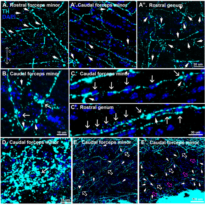
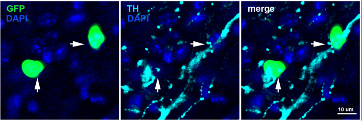
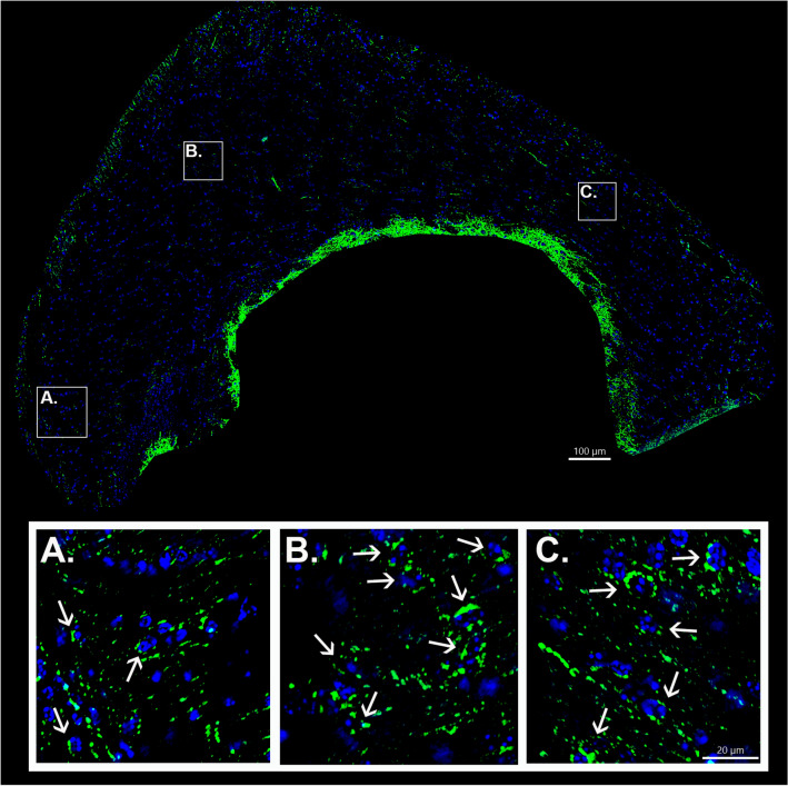
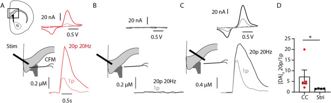
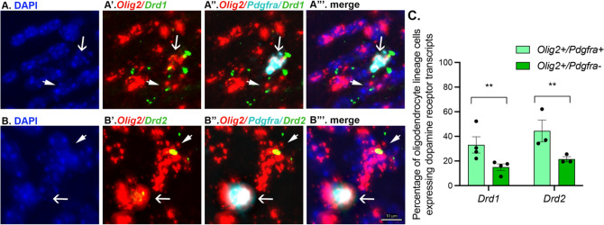
Oligodendrocyte progenitor cells (OPCs) receive synaptic innervation from glutamatergic and GABAergic axons and can be dynamically regulated by neural activity, resulting in activity-dependent changes in patterns of axon myelination. However, it remains unclear to what extent other types of neurons may innervate OPCs. Here, we provide evidence implicating midbrain dopamine neurons in the innervation of oligodendrocyte lineage cells in the anterior corpus callosum and nearby white matter tracts of male and female adult mice. Dopaminergic axon terminals were identified in the corpus callosum of DAT-Cre mice after injection of an eYFP reporter virus into the midbrain. Furthermore, fast-scan cyclic voltammetry revealed monoaminergic transients in the anterior corpus callosum, consistent with the anatomical findings. Using RNAscope, we further demonstrate that ~ 40% of Olig2 + /Pdfgra + cells and ~ 20% of Olig2 + /Pdgfra- cells in the anterior corpus callosum express Drd1 and Drd2 transcripts. These results suggest that oligodendrocyte lineage cells may respond to dopamine released from midbrain dopamine axons, which could affect myelination. Together, this work broadens our understanding of neuron-glia interactions with important implications for myelin plasticity by identifying midbrain dopamine axons as a potential regulator of corpus callosal oligodendrocyte lineage cells.
Keywords: Dopamine d1 receptor; Dopamine d2 receptor; Dopamine-glial interactions; Myelin plasticity; Oligodendrocyte progenitor cells.
© 2023. The Author(s).
Conflict of interest statement
The authors declare no competing interests.
Figures
References
-
- Albertson DN, Pruetz B, Schmidt CJ, Kuhn DM, Kapatos G, Bannon MJ (2004) Gene expression profile of the nucleus accumbens of human cocaine abusers: evidence for dysregulation of myelin. J Neurochem 88:1211–1219. https://www.ncbi.nlm.nih.gov/pmc/articles/PMC2215309/ - PMC - PubMed
-
- Anderegg A, Poulin J-F, Awatramani R (2015) Molecular heterogeneity of midbrain dopaminergic neurons—moving toward single cell resolution. FEBS Lett 589:3714–3726. 10.1016/j.febslet.2015.10.022(http://doi.wiley.com/) - PMC - PubMed
-
- Bacmeister CM, Barr HJ, McClain CR, Thornton MA, Nettles D, Welle CG, Hughes EG (2020) Motor learning promotes remyelination via new and surviving oligodendrocytes. Nat Neurosci 23:819–831. http://www.nature.com/articles/s41593-020-0637-3 - PMC - PubMed
-
- Beeler JA, Kisbye Dreyer J (2019) Synchronicity: the role of midbrain dopamine in whole-brain coordination. eNeuro 6:ENEURO.0345–18.2019. 10.1523/ENEURO.0345-18.2019(https://www.eneuro.org/lookup/doi/) - PMC - PubMed
-
- Bongarzone ER, Howard SG, Schonmann V, Campagnoni AT (1998) Identification of the dopamine D3 receptor in oligodendrocyte precursors: potential role in regulating differentiation and myelin formation. J Neurosci 18:5344–5353. 10.1523/JNEUROSCI.18-14-05344.1998(http://www.jneurosci.org/lookup/doi/) - PMC - PubMed
MeSH terms
Substances
Grants and funding
LinkOut - more resources
Full Text Sources
Miscellaneous
