

A novel approach to determining augmented bone volume in intraoral bone block augmentation using an intraoral scanner: an in vitro study
- PMID: 37668754
- PMCID: PMC10480375
- DOI: 10.1186/s40729-023-00492-0
A novel approach to determining augmented bone volume in intraoral bone block augmentation using an intraoral scanner: an in vitro study
Abstract
Introduction: Bone augmentation procedures are established tools for reshaping the alveolar ridge and increasing bone volume. Different approaches are being used to measure postoperative bone volume gain. This study aimed to develop an objective and automated volume measurement tool equally as precise as manual slice-by-slice annotation.
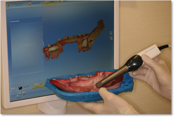
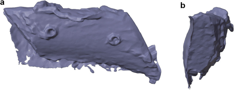
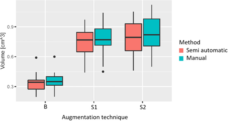
Materials and methods: To evaluate the proposed workflow, we performed an in vitro study with 20 pig mandibles that were grafted using three different grafting techniques-autogenous full block, split block bone and shell augmentation. The pig jaws were scanned pre- and postoperatively using an intraoral scanner. The resulting surface files (baseline, full block, split block, shell) were processed using the new volume-measuring workflow as well as using manual slice-by-slice annotation at baseline (t0) and at 6 months (t1) using the same population. Two TOSTs (Test of One-Sided Significance) and NHSTs (Null Hypothesis Significance Test) were used to compare the two workflows. The intra-rater reliability between t0 and t1 was determined using intraclass correlation coefficients.
Results: The mean difference for the full block augmentation technique was - 0.015 cm3 (p < 0.001); for the split block technique, it was - 0.034 cm3 p = 0.01, and for the shell technique, it was - 0.042 cm3. All results were statistically not different from zero and statistically equivalent to zero. The results also showed an excellent absolute intra-rater agreement.
Conclusions: The semiautomatic volume measurement established in this article achieves comparable results to manual slice-by-slice measuring in determining volumes on STL files generated by intraoral scanners and shows an excellent intra-rater reliability.
Keywords: Autogenous bone; Automated volume measurement; Bone augmentation; Bone blocks, intraoral bone graft; Bone regeneration.
© 2023. Deutsche Gesellschaft für Implantologie im Zahn‐, Mund‐ und Kieferbereich e.V., Japanese Society of Oral Implantology.
Conflict of interest statement
The authors declare that they have no competing interests.
Figures


Similar articles
-
The Influence of Different Graft Designs of Intraoral Bone Blocks on Volume Gain in Bone Augmentation Procedures: An In Vitro Study.Int J Oral Maxillofac Implants. 2020 Nov/Dec;35(6):1083-1089. doi: 10.11607/jomi.8368. Int J Oral Maxillofac Implants. 2020. PMID: 33270046
-
The Effect of Membrane Coverage on the Resorption of Autogenous Intraoral Block Grafts in Horizontal Ridge Augmentation: A Systematic Review of Literature and Meta-Analysis: Inevitability or an Iatrogenic Vulnerability?J Evid Based Dent Pract. 2018 Dec;18(4):275-289. doi: 10.1016/j.jebdp.2017.11.001. Epub 2017 Nov 21. J Evid Based Dent Pract. 2018. PMID: 30514442
-
Fully digital versus conventional workflow for horizontal ridge augmentation with intraoral block bone: A randomized controlled clinical trial.Clin Implant Dent Relat Res. 2022 Dec;24(6):809-820. doi: 10.1111/cid.13129. Epub 2022 Sep 6. Clin Implant Dent Relat Res. 2022. PMID: 36068075 Clinical Trial.
-
3D vertical alveolar crest augmentation in the posterior mandible using the tunnel technique: A 10-year clinical study.Int J Oral Implantol (Berl). 2022 May 13;15(2):111-126. Int J Oral Implantol (Berl). 2022. PMID: 35546722
-
Clinical performance of alveolar ridge augmentation with xenogeneic bone block grafts versus autogenous bone block grafts. A systematic review.J Stomatol Oral Maxillofac Surg. 2021 Jun;122(3):293-302. doi: 10.1016/j.jormas.2020.10.009. Epub 2020 Nov 5. J Stomatol Oral Maxillofac Surg. 2021. PMID: 33161168
References
-
- Jensen SS, Terheyden H. Bone augmentation procedures in localized defects in the alveolar ridge: clinical results with different bone grafts and bone-substitute materials. Int J Oral Maxillofac Implants. 2009;24(Suppl):218–236. - PubMed
-
- Acocella A, Bertolai R, Colafranceschi M, Sacco R. Clinical, histological and histomorphometric evaluation of the healing of mandibular ramus bone block grafts for alveolar ridge augmentation before implant placement. J Craniomaxillofac Surg. 2010;38(3):222–230. doi: 10.1016/j.jcms.2009.07.004. - DOI - PubMed
Publication types
MeSH terms
LinkOut - more resources
Full Text Sources
