

Impact of brain segmentation methods on regional metabolism quantification in 18F-FDG PET/MR analysis
- PMID: 37668814
- PMCID: PMC10480127
- DOI: 10.1186/s13550-023-01028-8
Impact of brain segmentation methods on regional metabolism quantification in 18F-FDG PET/MR analysis
Abstract
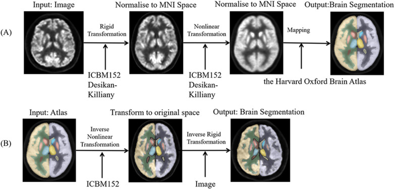
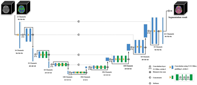
Background: Accurate analysis of quantitative PET data plays a crucial role in studying small, specific brain structures. The integration of PET and MRI through an integrated PET/MR system presents an opportunity to leverage the benefits of precisely aligned structural MRI and molecular PET images in both spatial and temporal dimensions. However, in many clinical workflows, PET studies are often performed without the aid of individually matched structural MRI scans, primarily for the sake of convenience in the data collection and brain segmentation possesses. Currently, two commonly employed segmentation strategies for brain PET analysis are distinguished: methods with or without MRI registration and methods employing either atlas-based or individual-based algorithms. Moreover, the development of artificial intelligence (AI)-assisted methods for predicting brain segmentation holds promise but requires further validation of their efficiency and accuracy for clinical applications. This study aims to compare and evaluate the correlations, consistencies, and differences among the above-mentioned brain segmentation strategies in quantification of brain metabolism in 18F-FDG PET/MR analysis.
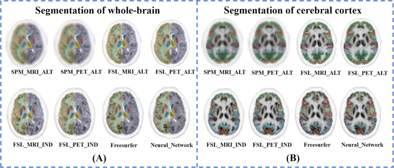
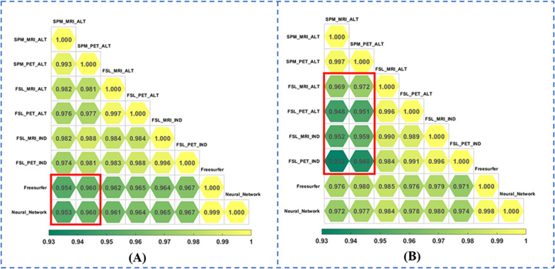
Results: Strong correlations were observed among all methods (r = 0.932 to 0.999, P < 0.001). The variances attributable to subject and brain region were higher than those caused by segmentation methods (P < 0.001). However, intraclass correlation coefficient (ICC)s between methods with or without MRI registration ranged from 0.924 to 0.975, while ICCs between methods with atlas- or individual-based algorithms ranged from 0.741 to 0.879. Brain regions exhibiting significant standardized uptake values (SUV) differences due to segmentation methods were the basal ganglia nuclei (maximum to 11.50 ± 4.67%), and various cerebral cortexes in temporal and occipital regions (maximum to 18.03 ± 5.52%). The AI-based method demonstrated high correlation (r = 0.998 and 0.999, P < 0.001) and ICC (0.998 and 0.997) with FreeSurfer, substantially reducing the time from 8.13 h to 57 s on per subject.
Conclusions: Different segmentation methods may have impact on the calculation of brain metabolism in basal ganglia nuclei and specific cerebral cortexes. The AI-based approach offers improved efficiency and is recommended for its enhanced performance.
Keywords: Artificial intelligence; Magnetic resonance imaging; Metabolism; Positron emission tomography.
© 2023. Springer-Verlag GmbH Germany, part of Springer Nature.
Conflict of interest statement
The authors have no relevant financial or non-financial interests to disclose.
Figures
Similar articles
-
Quantitative analysis of MRI-guided attenuation correction techniques in time-of-flight brain PET/MRI.Neuroimage. 2016 Apr 15;130:123-133. doi: 10.1016/j.neuroimage.2016.01.060. Epub 2016 Feb 4. Neuroimage. 2016. PMID: 26853602
-
A computational pipeline for quantification of pulmonary infections in small animal models using serial PET-CT imaging.EJNMMI Res. 2013 Jul 23;3(1):55. doi: 10.1186/2191-219X-3-55. EJNMMI Res. 2013. PMID: 23879987 Free PMC article.
-
Fast and Accurate Amyloid Brain PET Quantification Without MRI Using Deep Neural Networks.J Nucl Med. 2023 Apr;64(4):659-666. doi: 10.2967/jnumed.122.264414. Epub 2022 Nov 3. J Nucl Med. 2023. PMID: 36328490 Free PMC article.
-
Regional SUV quantification in hybrid PET/MR, a comparison of two atlas-based automatic brain segmentation methods.EJNMMI Res. 2020 Jun 8;10(1):60. doi: 10.1186/s13550-020-00648-8. EJNMMI Res. 2020. PMID: 32514906 Free PMC article.
-
A multi-atlas based method for automated anatomical Macaca fascicularis brain MRI segmentation and PET kinetic extraction.Neuroimage. 2013 Aug 15;77:26-43. doi: 10.1016/j.neuroimage.2013.03.029. Epub 2013 Mar 26. Neuroimage. 2013. PMID: 23537938
References
-
- Cui B, Zhang T, Ma Y, Chen Z, Ma J, Ma L, et al. Simultaneous PET/MRI imaging of cerebral blood flow and glucose metabolism in the symptomatic unilateral internal carotid artery/middle cerebral artery steno-occlusive disease. Eur J Nucl Med Mol Imaging. 2020;47:1668–1677. doi: 10.1007/s00259-019-04551-w. - DOI - PMC - PubMed
-
- Song S, Cheng Y, Ma J, Wang L, Dong C, Wei Y, et al. Simultaneous FET-PET and contrast-enhanced MRI based on hybrid PET/MR improves delineation of tumor spatial biodistribution in gliomas: a biopsy validation study. Eur J Nucl Med Mol Imaging. 2020;47:1458–1467. doi: 10.1007/s00259-019-04656-2. - DOI - PMC - PubMed
-
- Guo K, Wang J, Wang Z, Wang Y, Cui B, Zhao G, et al. Morphometric analysis program and quantitative positron emission tomography in presurgical localization in MRI-negative epilepsies: a simultaneous PET/MRI study. Eur J Nucl Med Mol Imaging. 2022;49(6):1930–1938. doi: 10.1007/s00259-021-05657-w. - DOI - PubMed
Grants and funding
LinkOut - more resources
Full Text Sources
