Multicenter Study
. 2023 Oct 1;80(10):1105-1112.
doi: 10.1001/jamaneurol.2023.2974.

Rapid Immunodot AQP4 Assay for Neuromyelitis Optica Spectrum Disorder

Affiliations
Multicenter Study

Rapid Immunodot AQP4 Assay for Neuromyelitis Optica Spectrum Disorder

Ying Fu et al. JAMA Neurol. .

Abstract

Importance: Immunoglobulin G autoantibodies for aquaporin 4 (AQP4-IgG) serve as diagnostic biomarkers for neuromyelitis optica spectrum disorder (NMOSD), and the most sensitive and specific laboratory tests for their detection are cell-based assays (CBAs). Nevertheless, the limited availability of special instruments limits the widespread use of CBAs in routine laboratories.

Objective: To validate an enzyme immunodot assay for simple and rapid detection of AQP4-IgG.

Design, setting, and participants: This multicenter case-control study, conducted from May 2020 to February 2023, involved 4 medical centers (3 in China and 1 in Korea). The study included patients with AQP4-IgG-positive NMOSD, patients with other immune-related diseases, and healthy control individuals. Participants were excluded if they did not agree to participate or if their serum sample had turbidity.

Exposures: Serum AQP4 antibodies measured with immunodot assay.

Main outcomes and measures: The main outcome was performance of the immunodot assay compared with the gold standard CBA for detecting AQP4-IgG. To examine generalizability, cross-validation in Korea and at a second site in China, validation of patients with other immune-related diseases, and follow-up validation of the original cohort were performed.

Results: A total of 836 serum samples were collected; 400 were included in the diagnostic study and 436 in the validation sets. In a head-to-head diagnostic study involving 200 patients with NMOSD with AQP4-IgG (mean [SD] age, 43.1 [13.5] years; 188 [94%] female) and 200 healthy controls, use of an immunodot assay demonstrated antibody detection performance comparable to that of the gold standard (κ = 98.0%). The validation sets included 47 patients with NMOSD and 26 patients with other autoimmune diseases from Korea, 31 patients with NMOSD at a second site in China, 275 patients with other diseases, and 57 patients with NMOSD at follow-up. In the validation study, of 436 cases, 2 (<1%) were false positive and none were false negative. The CBA identified 332 AQP4-IgG-positive samples and 504 negative samples (200 [40%] in controls and 304 [60%] in patients with other diseases); 2 of the positive cases (<1%) were false negative and 4 of the negative cases (<1%) were false positive. The overall sensitivity of the immunodot assay was 99.4% (95% CI, 97.8%-99.9%), and the specificity was 99.2% (95% CI, 98.0%-99.8%).

Conclusions and relevance: This case-control study found that the immunodot assay was comparable to CBA for detecting AQP4-IgG. With its time- and cost-efficient characteristics, the immunodot assay may be a practical option for AQP4-IgG detection.


Conflict of interest statement

Conflict of Interest Disclosures: Dr H. J. Kim reported receiving grants from the National Research Foundation of Korea; receiving personal fees from APRILBIO and Eisai; receiving consulting or speaker fees from or serving on a steering or scientific committee for Alexion, APRILBIO, ALTOS Biologics, Biogen, Celltrion, Daewoong, Eisai, GC Pharma, Handok, Horizon Therapeutics (formerly Viela Bio), Kaigene, Kolon Life Science, MDimune, Mitsubishi Tanabe Pharma, Merck Serono, Novartis, Roche, Sanofi Genzyme, Teva-Handok, and UCB outside the submitted work; and serving as coeditor for the Multiple Sclerosis Journal and associate editor for the Journal of Clinical Neurology. No other disclosures were reported.

Figures

Figure 1.
Figure 1.. Enzyme Immunodot Assay for Detection of Immunoglobulin G Autoantibodies Against Aquaporin 4 (AQP4-IgG)
B, For dual staining, patient serum and rabbit monoclonal AQP4-specific IgG were applied to the plasma membrane of cultured AQP4-expressing stable human embryonic kidney 293T (HEK-293T) cells. Secondary antibodies were used for detection. Green indicates Alexa Fluor 488–conjugated anti–human IgG and red, Alexa Fluor 594-conjugated goat anti–rabbit IgG. For patients, AQP4-IgG–positive patient serum is shown in green, AQP4-specific IgG in red, and merged images in yellow. For controls, AQP4-IgG–negative patient serum is shown in green, AQP4-specific IgG in red, and merged images in yellow. Scale bar, 50 μm. C, The contrast diagram shows the AQP4-IgG staining of patient samples with different titers. Cell-based assay results in HEK-293T cells expressing AQP4 are shown on the left, with enlarged views in the upper right corners; immunodot testing results for the same sample are shown on the right. Scale bar, 100 μm. NC indicates nitrocellulose filter and OAP, orthogonal array of particles.
Figure 2.
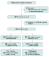
Figure 2.. Patient Flow Diagram and Results From the Diagnostic Study
Turbidity was defined as samples that appeared unclear with both the naked eye and against a lighted background. AQP4-IgG indicates immunoglobulin G autoantibodies against aquaporin 4 and CBA, cell-based assay.
Figure 3.
Figure 3.. International Cross-Validation in Korea
AQP4-IgG indicates immunoglobulin G autoantibodies against aquaporin 4; CBA, cell-based assay; and MOG-IgG, myelin oligodendrocyte glycoprotein antibodies.

Comment in

References

    1. O’Riordan JI, Gallagher HL, Thompson AJ, et al. Clinical, CSF, and MRI findings in Devic’s neuromyelitis optica. J Neurol Neurosurg Psychiatry. 1996;60(4):382-387. doi: 10.1136/jnnp.60.4.382 - DOI - PMC - PubMed
    1. Wingerchuk DM, Hogancamp WF, O’Brien PC, Weinshenker BG. The clinical course of neuromyelitis optica (Devic’s syndrome). Neurology. 1999;53(5):1107-1114. doi: 10.1212/WNL.53.5.1107 - DOI - PubMed
    1. Wingerchuk DM, Banwell B, Bennett JL, et al. ; International Panel for NMO Diagnosis . International consensus diagnostic criteria for neuromyelitis optica spectrum disorders. Neurology. 2015;85(2):177-189. doi: 10.1212/WNL.0000000000001729 - DOI - PMC - PubMed
    1. Pittock SJ, Zekeridou A, Weinshenker BG. Hope for patients with neuromyelitis optica spectrum disorders—from mechanisms to trials. Nat Rev Neurol. 2021;17(12):759-773. doi: 10.1038/s41582-021-00568-8 - DOI - PubMed
    1. Jarius S, Paul F, Weinshenker BG, Levy M, Kim HJ, Wildemann B. Neuromyelitis optica. Nat Rev Dis Primers. 2020;6(1):85. doi: 10.1038/s41572-020-0214-9 - DOI - PubMed

Publication types