. 2023 Oct 9;8(19):e167215.
doi: 10.1172/jci.insight.167215.

Tregs integrate native and CAR-mediated costimulatory signals for control of allograft rejection

Affiliations

Tregs integrate native and CAR-mediated costimulatory signals for control of allograft rejection

Isaac Rosado-Sánchez et al. JCI Insight. .

Abstract

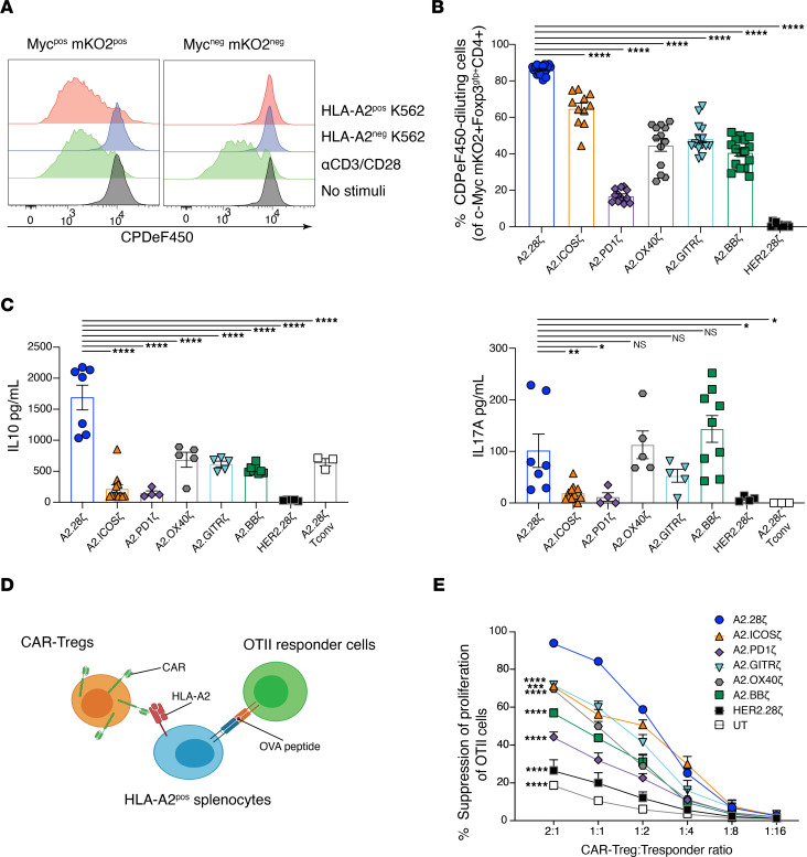
Tregs expressing chimeric antigen receptors (CAR-Tregs) are a promising tool to promote transplant tolerance. The relationship between CAR structure and Treg function was studied in xenogeneic, immunodeficient mice, revealing advantages of CD28-encoding CARs. However, these models could underrepresent interactions between CAR-Tregs, antigen-presenting cells (APCs), and donor-specific Abs. We generated Tregs expressing HLA-A2-specific CARs with different costimulatory domains and compared their function in vitro and in vivo using an immunocompetent model of transplantation. In vitro, the CD28-encoding CAR had superior antigen-specific suppression, proliferation, and cytokine production. In contrast, in vivo, Tregs expressing CARs encoding CD28, ICOS, programmed cell death 1, and GITR, but not 4-1BB or OX40, all extended skin allograft survival. To reconcile in vitro and in vivo data, we analyzed effects of a CAR encoding CD3ζ but no costimulatory domain. These data revealed that exogenous costimulation from APCs can compensate for the lack of a CAR-encoded CD28 domain. Thus, Tregs expressing a CAR with or without CD28 are functionally equivalent in vivo, mediating similar extension of skin allograft survival and controlling the generation of anti-HLA-A2 alloantibodies. This study reveals a dimension of CAR-Treg biology and has important implications for the design of CARs for clinical use in Tregs.

Keywords: Costimulation; Immunology; Immunotherapy; Tolerance; Transplantation.


Conflict of interest statement

Conflict of interest: MKL received research funding from Bristol Myers Squibb. MKL is an inventor on pending patents (PCT/CA2028/051167 and PCT/CA2018/051174) related to HLA-A2–specific chimeric antigen receptors.

Figures

Figure 1
Figure 1. Design and expression of costimulatory domain CAR variants.
(A) Schematic diagram summarizing the TM and signaling domains incorporated into the second-generation CAR variants. (B) Representative flow cytometry plots of at least 3 independent experiments showing CAR (c-Myc) and mKO2 reporter expression and binding to an HLA-A2 tetramer, with percentages shown in corners. (C) MFI of CAR expression for different CAR variants in Tregs after expansion gated on live c-Myc+CD4+Foxp3gfp+ cells; n = 6–13 replicates from at least 8 independent experiments. (D) Foxp3gfp expression in Tregs after expansion, gated on live CD4+ cells; n = 6–16 replicates from at least 11 independent experiments. (E) Representative data of at least 5 independent experiments showing intracellular Foxp3 and Helios expression in CAR-Tregs and control conventional T cells (Tconv) after expansion, gated on total live CD4+ cells. Data reported as mean ± SEM. Statistical significance was determined using 1-way ANOVA with a Holm-Šidak posttest. **P < 0.01, ***P < 0.001, ****P < 0.0001. Co-stim, costimulatory; UT, untransduced.
Figure 2
Figure 2. Costimulatory CAR variants differ in their ability to stimulate Tregs.
(AC) Tregs expressing the indicated CAR were stained with CPDe450 and cocultured with HLA-A2+ or HLA-A2 K562 cells, polyclonal stimulated with anti-CD3/28, or left unstimulated for 3 days. (A) Representative histograms of at least 5 independent experiments comparing A2.28ζ CAR-Treg proliferation of gated CAR+ (c-Myc+mKO2+) or CAR (c-MycmKO2) cells. (B) Frequencies of CAR-Tregs that divided after 3 days of coculture with HLA-A2+ K562s, determined by CPDeF450 dilution, gated on c-Myc+mKO2+Foxp3gfp+CD4+ cells; n = 11–20 replicates from at least 5 independent experiments. (C) Cytokine secretion after 3 days of coculture with HLA-A2+ K562s; n = 3–12 replicates from at least 3 independent experiments. (D and E) CAR-Tregs were cocultured with OTII CD4+ T cells at varying ratios in the presence of irradiated HLA-A2+ splenocytes and OVA peptide. (D) Schematic diagram of the linked suppression assay. (E) CAR-Treg–mediated suppression of the OTII CD4+ T cell proliferation, as determined by Ki67 expression; n = 3–6 replicates from at least 2 independent experiments. Tresponder, responder T cell; UT, untransduced. Data are reported as mean ± SEM. Statistical significance was determined using 1-way (B and C) or 2-way (E) ANOVA with a Holm-Šidak posttest comparing to CD28-based CAR-Tregs. *P < 0.05, **P < 0.01, ***P < 0.001, ****P < 0.0001.
Figure 3
Figure 3. In vivo effects of Tregs expressing costimulatory CAR variants on skin rejection.
BL/6 mice were transplanted with skin grafts from syngeneic or HLA-A2+ BL/6 mice and administered 1 × 10⁶ CAR-Tregs i.v. (A) Skin graft survival curves and (B) levels of anti–HLA-A2 IgG Abs from mice infused with Tregs expressing CARs encoding costimulatory domains from CD28 (left) or TNFR (right) family members. (A) Data from mice receiving no Treg treatment (PBS) or transplanted with syngeneic WT BL/6 grafts are shown in both graphs. (C) Correlation between anti–HLA-A2 IgG Abs in plasma at day 14 and skin graft rejection of mice receiving Tregs bearing CD28 family–based CARs. (D) Correlation between anti–HLA-A2 IgG Abs in plasma at day 14 and skin graft rejection of mice receiving Tregs bearing TNFR family–based CARs. (E) Persistence of CAR-Tregs measured as the percentage of Thy1.1+ CAR-Tregs of total CD45+ T cells in peripheral blood over time. (F and G) Phenotype of Thy1.1+CD4+ CAR-Tregs in peripheral blood over time including expression of (F) CAR (c-Myc+) and (G), FoxP3 alone (left), and FoxP3 with Helios (right). Data are reported as mean ± SEM pooled from 4 individual experiments with n = 3–13 mice per group. Statistical significance was determined using (A) log-rank Mantel-Cox test, (B and EG) 2-way ANOVA with a Holm-Šidak posttest, and Pearson’s correlation (C and D). *P < 0.05, **P < 0.01, ****P < 0.0001.
Figure 4
Figure 4. A CAR costimulatory domain is dispensable for CAR-Tregs in vivo.
(A) Schematic diagram of the first- and second-generation CARs used. Tregs expressing the indicated CARs were stained with CPDe450 and cocultured with HLA-A2+ K562 cells for 3 days. (B) The percentage of CAR-Tregs that divided, determined by CPDeF450 dilution (left); n = 12–20 replicates from at least 5 independent experiments; and IL-10 secretion (right), measured in culture supernatants; n = 5–7 replicates from at least 3 independent experiments. (C) CAR-Tregs were cocultured with OTII CD4+ T cells at varying ratios in the presence of irradiated HLA-A2+ splenocytes and OVA peptide. CAR-Tregs mediated suppression of the OTII CD4+ T cell proliferation, as determined by Ki67 expression; n = 3–6 replicates from at least 2 independent experiments. UT, untransduced. (DH) BL/6 mice were transplanted with skin grafts from syngeneic or HLA-A2+ BL/6 mice and administered 1 × 10⁶ CAR-Tregs i.v. (D) Skin graft survival curves and (E) levels of anti–HLA-A2 IgG Abs from mice infused with Tregs expressing first- and second-generation CARs. (F) Persistence of CAR-Tregs measured as the percentage of Thy1.1+ CAR-Tregs of total CD45+ T cells in peripheral blood over time. (G and H) CAR-Treg expression of (G) CAR (c-Myc+) and (H) FoxP3 and FoxP3 and Helios. In vivo data pooled from 3 individual experiments with n = 3–13 mice per group. Data are reported as mean ± SEM. Data from the A2.28z, HER2.28z, and UT conditions are also shown in Figure 2, C and E, and Figure 3, A, B, and E–G. Statistical significance was determined using (B) 1-way or (C and EH) 2-way ANOVA with a (C and E) Holm-Šidak posttest or (D) log-rank Mantel-Cox test. **P < 0.01, ***P < 0.001, ****P < 0.0001. Co-stim, costimulatory; UT, untransduced.
Figure 5
Figure 5. Effect of exogenous costimulation on CAR-Tregs.
CAR-Tregs were co-cultured with CD86posHLA-A2pos or CD86negHLA-A2pos K562 cells at a 1:2 K562/Tregs ratio for 3 days. (A) Schematic diagram of assay. (B and C) Ki67 and CTLA-4 expression in CAR-Tregs following 3 days of co-culture, gated on c-Myc+CD4+ live cells; n = 4 to 6 replicates from 2 independent experiments. (D) LAP expression in different costimulatory-encoding CAR-Tregs following 3-days of co-culture, gated on c-Myc+CD4+ live cells; n = 2 to 3 replicates from 1 experiment. (E) Schematic diagram of the CD8α-TM CARs generated. (F) Ki67 and LAP expression in first-generation CD8α-TM CAR-Tregs following 3 days of co-culture, gated on c-Myc+CD4+ live cells; n = 4 to 6 replicates from 1 experiment. Where indicated, CTLA-4–Ig and an anti-CD28 agonist mAb were added at 10 μg/mL. Data are reported as mean ± SEM. Statistical significance was determined using 1-way (C and F) or 2-way (B and D) ANOVA with a Holm-Šidak posttest. *P < 0.05, **P < 0.01, ***P < 0.001, ****P < 0.0001. Co-stim, costimulatory.
Figure 6
Figure 6. In vivo and in vitro APC suppression by first- and second-generation CAR-Tregs.
(AD and G) For in vitro assays, CAR-Tregs were cocultured with splenic HLA-A2+ CD11c+ DCs at a ratio of 1:2 or 1:5 DCs to Tregs for 1 or 2 days. (A) Schematic of in vitro DC suppression assay. (B) Representative histograms of at least 5 independent experiments showing CD80 expression on CD11c+ DCs after 2-days of culture with the indicated types of Tregs. (C) Expression of CD80 (left) and CD86 (right) in HLA-A2+ CD11c+ DCs relative to DCs cultured with untransduced Tregs (dotted line); n = 8–15 replicates from at least 5 independent experiments. (D) In vitro DC suppression assays performed with or without 10 μg/mL CTLA-4–Ig; n = 3 replicates from 2 independent experiments. (E and F) For in vivo assays, BL/6 mice were transplanted with skin grafts from HLA-A2+ BL/6 mice and treated or not with 1 × 10⁶ CAR-Tregs. dLN and spleen tissues were collected at day 7 after CAR-Treg infusion; n = 6–7 mice per group from 2 independent experiments. (E) Expression of CD80, CD86, and MHC-II in DCs from dLNs of mice treated with CAR-Tregs relative to untreated mice (dotted lines; average of 3–4 untreated mice per each experiment). (F) Expression of CD80, CD86, and MHC-II in DCs from spleens of mice treated with CAR-Tregs relative to untreated mice (dotted lines; average of 3–4 untreated mice per each experiment). (G) Expression of CD80 at day 1 (left), CD86 at day 2 (center), and MHC-II at day 1 (right) on HLA-A2+CD11c+ DCs treated with different types of CAR-Tregs in vitro; n = 6–16 replicates from 4 (CD80/MHC-II) or 5 (CD86) independent experiments. (C, D, and G) Data are shown relative to DCs cultured with untransduced Tregs, which were normalized to 100% (dotted lines). (E and F) Data are shown relative to the expression of CD80, CD86, and MHC-II in DCs from nontreated mice, which were normalized to 100% (dotted lines). Data are reported as mean ± SEM. Statistical significance was determined using (C, D, and G) 1-way ANOVA with a Holm-Šidak posttest and (E and F) Student’s t test. *P < 0.05, **P < 0.01, ***P < 0.001, ****P < 0.0001.

References

    1. Lam AJ, et al. Harnessing advances in T regulatory cell biology for cellular therapy in transplantation. Transplantation. 2017;101(10):2277–2287. doi: 10.1097/TP.0000000000001757. - DOI - PubMed
    1. Raffin C, et al. Treg cell-based therapies: challenges and perspectives. Nat Rev Immunol. 2020;20(3):158–172. doi: 10.1038/s41577-019-0232-6. - DOI - PMC - PubMed
    1. Ferreira LMR, et al. Next-generation regulatory T cell therapy. Nat Rev Drug Discov. 2019;18(10):749–769. doi: 10.1038/s41573-019-0041-4. - DOI - PMC - PubMed
    1. Sawitzki B, et al. Regulatory cell therapy in kidney transplantation (the ONE study): a harmonised design and analysis of seven non-randomised, single-arm, phase 1/2A trials. Lancet. 2020;395(10237):1627–1639. doi: 10.1016/S0140-6736(20)30167-7. - DOI - PMC - PubMed
    1. Harden PN, et al. Feasibility, long-term safety, and immune monitoring of regulatory T cell therapy in living donor kidney transplant recipients. Am J Transplant. 2021;21(4):1603–1611. doi: 10.1111/ajt.16395. - DOI - PMC - PubMed

Publication types