. 2023 Sep 5;19(9):e1011632.
doi: 10.1371/journal.ppat.1011632. eCollection 2023 Sep.

Evidence for preexisting prion substrain diversity in a biologically cloned prion strain

Affiliations

Evidence for preexisting prion substrain diversity in a biologically cloned prion strain

Tess Gunnels et al. PLoS Pathog. .

Abstract

Prion diseases are a group of inevitably fatal neurodegenerative disorders affecting numerous mammalian species, including Sapiens. Prions are composed of PrPSc, the disease specific conformation of the host encoded prion protein. Prion strains are operationally defined as a heritable phenotype of disease under controlled transmission conditions. Treatment of rodents with anti-prion drugs results in the emergence of drug-resistant prion strains and suggest that prion strains are comprised of a dominant strain and substrains. While much experimental evidence is consistent with this hypothesis, direct observation of substrains has not been observed. Here we show that replication of the dominant strain is required for suppression of a substrain. Based on this observation we reasoned that selective reduction of the dominant strain may allow for emergence of substrains. Using a combination of biochemical methods to selectively reduce drowsy (DY) PrPSc from biologically-cloned DY transmissible mink encephalopathy (TME)-infected brain resulted in the emergence of strains with different properties than DY TME. The selection methods did not occur during prion formation, suggesting the substrains identified preexisted in the DY TME-infected brain. We show that DY TME is biologically stable, even under conditions of serial passage at high titer that can lead to strain breakdown. Substrains therefore can exist under conditions where the dominant strain does not allow for substrain emergence suggesting that substrains are a common feature of prions. This observation has mechanistic implications for prion strain evolution, drug resistance and interspecies transmission.


Conflict of interest statement

The authors have declared that no competing interests exist.

Figures

Fig 1
Fig 1. Suppression of HY PrPSc formation by DY TME.
(A-C) Western blot analysis of representative PMCA serial dilution samples (n = 3) of DY PrPSc alone (A), HY PrPSc alone (B) and mixtures of HY and DY PrPSc (C). Input samples were ten-fold serially diluted, subjected to one round of PMCA followed by PK digestion, and probed by immunoblotting using antibodies 3F4 (detects all strains) and 12B2 (specific for an epitope present on HY PrPSc but not DY PrPSc). Both strains amplify independently, with HY PrPSc having higher replication efficiency compared to DY PrPSc. When mixed at a constant HY to DY ratio of 1:1000, the 500:0.5 μg eq mixture shows complete suppression of HY PrPSc amplification, while the 50:0.05 μg eq. mixture shows incomplete suppression, with some HY PrPSc being detectable in the sample using the 12B2 antibody. When DY PrPSc is below 50 μg eq, HY PrPSc amplifies without interference.
Fig 2
Fig 2. Extended PK digestion of DY TME-infected brain homogenate reveals the presence of non-DY PrPSc species.
Western blot analysis of proteinase K strain selection assay products seeded with uninfected (UN; panel A,C,E,G) or drowsy (DY) brain homogenate (panels B, D, F, H) after one (panels A-D) or two (panels E-H) rounds of PMCA probed with either the anti-PrP antibody 3F4 (panels A-B, E-F) or 12B2 (panels C-D, G-H).
Fig 3
Fig 3. Detection of prion substrains in DY TME-infected brain.
Western blot analysis of conformational strain selection assay products seeded with uninfected (UN; panel A,B,E,F,I,J,M,N) or drowsy (DY) brain homogenate at either 2M (panels C,G,K,O) or 4M (panels D,H,L,P) Gdn-HCl after one (panels A-H) or two (panels I-P) rounds of PMCA probed with either the anti-PrP antibody 3F4 (panels A-D, I-L) or 12B2 (panels E-H, M-P).
Fig 4
Fig 4. Hamsters infected with CSSA products have a bona fide prion infection.
Western blot analysis of proteinase K digested brain homogenate from mock infected hamster (UN; lane 1), DY TME infected hamster (DY; lane 2), HY TME infected hamster (HY; lane 3) or CSSA products from mock-infected reactions (lanes 4 and 5) or DY CSSA reactions with either 2M (lane 6) or 4M (lane 7) Gdn-HCl. Second (lanes 8 and 9) and third (lane 10) serial hamster passage of brain material from hamsters infected with CSSA products from lanes 6 and 7. Western blots were probed with either the anti-PrP antibody 3F4 (top panel) that recognizes both the 19 (lane 2) and 21 kDa (lane 3) unglycosylated PrPSc polypeptide or the anti-PrP antibody 12B2 which recognizes the 21 kDa (lane 3) but not the 19 kDa (lane 2) unglycosylated PrPSc polypeptide. The migration of the 19 and 21 kDa unglycosylated PrPSc polypeptide are indicated at the left of the panel.
Fig 5
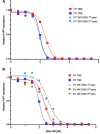
Fig 5. Conformational stability of PrPSc from hamsters infected with brain-derived prion strains and the CSSA isolated substrain differ.
Representative PrPSc conformational stability curves from hamsters infected with either HY TME, DY TME, 2M DY CSSA reaction products (panel A), or 4M DY CSSA reaction products (panel B). The conformational stability curves were repeated a minimum of 8 times with similar results.

Similar articles

Cited by

References

    1. Bruce ME, Will RG, Ironside JW, McConnell I, Drummond D, Suttie A, et al.. Transmissions to mice indicate that ’new variant’ CJD is caused by the BSE agent [see comments]. Nature. 1997;389(6650):498–501. - PubMed
    1. Fraser H, Bruce ME, Chree A, McConnell I, Wells GAH. Transmission of bovine spongiform encephalopathy and scrapie to mice. J Gen Virol. 1992;73:1891–7. doi: 10.1099/0022-1317-73-8-1891 - DOI - PubMed
    1. Wells GAH, Scott AC, Johnson CT, Gunning RF, Hancock RD, Jeffrey M, et al.. A novel progressive spongiform encephalopathy in cattle. Vet Rec. 1987;121:419–20. doi: 10.1136/vr.121.18.419 - DOI - PubMed
    1. Pritzkow S. Transmission, Strain Diversity, and Zoonotic Potential of Chronic Wasting Disease. Viruses. 2022;14(7). doi: 10.3390/v14071390 - DOI - PMC - PubMed
    1. Gilch S. Chronic wasting disease—A prion disease through a One Health lens. Can Vet J. 2022;63(4):431–3. - PMC - PubMed

Publication types