. 2023 Sep 5;14(1):5398.
doi: 10.1038/s41467-023-41039-6.

Notch and retinoic acid signals regulate macrophage formation from endocardium downstream of Nkx2-5

Affiliations

Notch and retinoic acid signals regulate macrophage formation from endocardium downstream of Nkx2-5

Norika Liu et al. Nat Commun. .

Abstract

Hematopoietic progenitors are enriched in the endocardial cushion and contribute, in a Nkx2-5-dependent manner, to tissue macrophages required for the remodeling of cardiac valves and septa. However, little is known about the molecular mechanism of endocardial-hematopoietic transition. In the current study, we identified the regulatory network of endocardial hematopoiesis. Signal network analysis from scRNA-seq datasets revealed that genes in Notch and retinoic acid (RA) signaling are significantly downregulated in Nkx2-5-null endocardial cells. In vivo and ex vivo analyses validate that the Nkx2-5-Notch axis is essential for the generation of both hemogenic and cushion endocardial cells, and the suppression of RA signaling via Dhrs3 expression plays important roles in further differentiation into macrophages. Genetic ablation study revealed that these macrophages are essential in cardiac valve remodeling. In summary, the study demonstrates that the Nkx2-5/Notch/RA signaling plays a pivotal role in macrophage differentiation from hematopoietic progenitors.


Conflict of interest statement

The authors declare no competing interests.

Figures
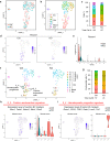
Fig. 1
Fig. 1. scRNA-seq analysis identified Nkx2-5-dependent cushion endocardial cells and hematopoietic progenitors.
a UMAP representation of merged dataset with cells color-coded by genotype (Ctrl or KO). b UMAP representation of single-cell gene expression showing the seven identified clusters. EC: endocardial cell, CM: cardiomyocyte, Blood: blood cell (leukocyte), Epi: pro-epicardium (c) Stacked bar plot showing the relative proportion of clusters in each genotype, with total cell counts indicated. d, e UMAP feature plots and violin plot of Pecam1 expression demonstrating that number of endocardial cells are decreased in KO. f UMAP representation of single-cell gene expression showing that the endocardial cluster can be subclustered into five (0_0 to 0_4) in Ctrl, but only three subclusters appear in KO. g Stacked bar plot showing the relative proportion of clusters in each genotype, with total cell counts indicated. h, i Module scores projected on the UMAP and violin plots. The signature plot demonstrating that cluster 0_3 was enriched for cushion endocardium marker genes (Msx1, Twist1, Notch1, Ifitm1, and Fbln2) (H) and cluster 0_4 was enriched for hematopoietic progenitor marker genes (Runx1, CD41(Itga2b), CD61 (Itgb3), Gata1, Lmo2, and Tal1) (I). Cell clusters of interest are indicated by * in the violin plot.
Fig. 2
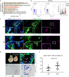
Fig. 2. Nkx2-5 regulation of Notch signaling is essential for endocardial hematopoiesis.
a Result from GO analysis from the differential expression genes (DEGs) between Ctrl and KO endocardium. b UMAP feature plots and violin plots showing the reduced expression level of Notch1 in KO endocardium. c Cushion endocardium of Ctrl and KO embryos stained for N1ICD (pink), CD31 (green), and DAPI (blue). N1ICD was stained in the cushion endocardial cells in the control, but fewer in the Nkx2-5-null embryos. Five experiments were repeated independently with similar results. scale: 50 μm. d Overexpression of NICD in Nkx2-5 lineage cells activates hematopoiesis. (i) Ctrl and Nkx2-5cre/+; R26NICD-GFP/+ embryos at E10.5. Although the mutants were smaller in body size, the size of the heart and the somite number were comparable. The heart was filled with blood (black arrow) and that head vasculatures are dilated and filled with blood as well (white arrow). (ii) Outflow tract of Nkx2-5cre/+; R26NICD-GFP/+ at E10.0. Many of the CD41+ cells in the outflow tract endocardium are costained with GFP reporter conjugated with N1ICD allele (white arrows). Many of the CD41+ cells are labeled with GFP reporter. (iii, iv) CD31 (red)/CD41 (blue) staining of control (iii) and Nkx2-5cre/+; R26NICD-GFP/+ (iv) embryos at E10.5 showed a striking increase in CD41+ cells (blue) in the mutant heart. e Flow cytometry analysis of the hearts from Ctrl and Nkx2-5cre/+; R26NICD-GFP/+ (Mut) embryos at E10.0. The number of CD31 + CD41+ hematopoietic progenitors is significantly increased in the Mut hearts. Data represent mean ± SEM. Ctrl: 12 embryos were analyzed from 4 litters (n = 12), Mut: 11 embryos were analyzed from 3 litters (n = 11), p = 0.0115 from unpaired, two-tailed t-test. Source data are provided as a Source Data file.
Fig. 3
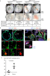
Fig. 3. Nkx2-5 mutant phenotype is rescued by overexpression of N1ICD.
a Whole embryos (i-iv) and enlarged images of hearts (v-viii). Knockout embryo showed pericardial effusion (black arrow head). Caudal part of the dorsal aorta and heart of both Overexpression and Rescue embryos were filled with blood (white arrow head). The total number of embryos analyzed and the number of those that exhibited phenotypes are indicated in the table below. b Sections of ventricle of hearts of Null and Rescue embryos at E10.5. Nkx2-5-null endocardium rarely expressed CD41 ((i)(i)’). However, N1ICD overexpression (Rescue) restored endocardial CD41+ cells, many of which are co-labeled with GFP ((ii)(ii)’). CD31 staining showed that Nkx2-5-null lacks cushion endocardium (iii), but the morphology of outflow tract of Rescue embryo appears normal with the presence of cushion endocardium (iv)(iv)’. c Quantification of CD41+ hematopoietic progenitors derived from Nkx2-5-lineage (GFP+) by immunofluorescence imaging on null and rescue heart. Data are presented as mean values +/− SEM. Representative sections (those with the highest number of positive cells) from 4 embryos were quantified (n = 4), p = 0.0033 from unpaired, two-tailed t-test. Source data are provided as a Source Data file.
Fig. 4
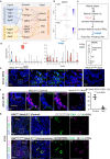
Fig. 4. Retinoic acid signaling is suppressed in endocardial hematopoiesis via Dhrs3 expression.
a Schematic presentation of ligand-receptor-target networks summarized from NicheNet analysis. Those with both receptors and targes are expressed in the hemogenic endocardium are shown. Arrows connecting ligands and receptors were predicted from the NicheNet packages as indicated in Supplementary Fig. 4c. Receptors and targets were connected (dashed arrows) based on the results of NicheNet ligand-target prediction as shown in Supplementary Fig. 4b. b UMAP feature plots showing Dhrs3 expression is highly enriched in hemogenic EC in Ctrl but absent in KO. c Simplified scheme of RA metabolites. d Violin plots showed that Dhrs3 is abundantly expressed and Crabp2 is deficient in hemogenic EC (0_4). e, f Sections of atriums of control and Nkx2-5-null embryos at E9.5. Dhrs3 was specifically expressed on CD41+ cells, as well as CD31+ endocardial cells. g Dhrs3+CD31+ cells in total CD31+ cells were quantified from immunofluorescent staining. Data are presented as mean values +/− SEM. Representative sections (those with the highest number of positive cells) from 5 embryos were quantified (n = 5), p < 0.0001 from unpaired, two-tailed t-test. Source data are provided as a Source Data file. h Forced activation of Notch signaling increases the number of Dhrs3+ macrophages in the cardiac cushion at E13.5 embryos. Double positive cells for CD68 and Dhrs3 are indicated with yellow arrow heads. Ratio of Dhrs3+ macrophages were 33% and 75% in Control and Overexpression embryos, respectively. Only one embryos could be obtained, but the similar results were reproduced using Nkx2-5cre/+; CAG-CAT-N1ICDtg mice, which are not lethal at E13.5. These data are shown in Supplementary Fig. 6b, c. PAV Pulmonary arterial valve.
Fig. 5
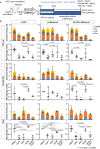
Fig. 5. Notch signaling promotes, whereas retinoic acid suppresses, macrophage differentiation.
a Schematic representation of the three regimens of the colony-forming assay using hematopoietic organs of embryos at pre-circulation stage. Enzymatically digested tissues were co-cultured with OP9 feeder cells for 4 days, followed by methylcellulose culture with or without atRA (active RA), DAPT (Notch inhibitor) or DEAB (RA synthase inhibitor). Colonies were counted by the light microscopy and further analyzed by flow cytometry for macrophage ratio. b The number of hematopoietic colonies obtained from individual experiments was normalized by the number of macrophage colonies from the DMSO treatment. Results of the macrophage colony were pulled out in the lower panel of each tissue. Both DAPT and atRA consistently reduced hematopoietic activity in regimen (i), but the effect was blunted and less consistent among tissue in regimen (ii). GM: granulocyte-macrophage, GEMM: granulocyte-erythroid-macrophage-megakaryocyte, Ery Erythrocyte, Mac Macrophage. Data are presented as mean values +/− SEM. Yolk sac: Four independent experiments with 1 or 2 replications in each condition were performed (n = 4). Caudal half: Four independent experiments (5 embryos were pooled for each experiment) in each condition were performed (n = 4). Heart: Three independent experiments (5 embryos were pooled for each experiment) with 1 or 2 replications in each condition were performed (n = 3). p values were calculated by One-way ANOVA with post-hoc Tukey’s multiple comparisons test. Source data are provided as a Source Data file.
Fig. 6
Fig. 6. Ablation of Csf1r (macrophage surface marker) in Nkx2-5 lineage results in cardiac enlargement, valvular degeneration, and narrow aortic root.
Nkx2-5cre/+; Csf1rflox/flox hearts and their controls (3 other genotypes) were dissected at 12 weeks old. Mice were all in C57Bl/6 background. a Mutants were born at normal Mendelian rate. b Appearance of whole heart. Note the enlargement of the heart. c Histology of aortic root and valves by Pentachrome staining. Valves show a spectrum of degenerations (light blue arrow). Blue line indicates the aortic root measured just above the aortic cusp. Three experiments were repeated independently and two of them showed the similar results. d Schematic summary of the study. Notch signaling induces macrophage differentiation of Nkx2-5+ hemogenic endocardial cells by suppressing retinoic acid signaling through Dhrs3 expression. Macrophages of Nkx2-5-lineage are required for valve maturation.

References

    1. Zhang H, Lui KO, Zhou B. Endocardial Cell Plasticity in Cardiac Development, Diseases and Regeneration. Circ. Res. 2018;122:774–789. doi: 10.1161/CIRCRESAHA.117.312136. - DOI - PubMed
    1. Nakano H, et al. Haemogenic endocardium contributes to transient definitive haematopoiesis. Nat. Commun. 2013;4:1564. doi: 10.1038/ncomms2569. - DOI - PMC - PubMed
    1. Lluri G, et al. Hematopoietic progenitors are required for proper development of coronary vasculature. J. Mol. Cell. Cardiol. 2015;86:199–207. doi: 10.1016/j.yjmcc.2015.07.021. - DOI - PMC - PubMed
    1. Yzaguirre AD, Speck NA. Insights into blood cell formation from hemogenic endothelium in lesser-known anatomic sites. Dev. Dyn. Publ. Am. Assoc. Anat. 2016;245:1011–1028. - PMC - PubMed
    1. Zamir, L. et al. Nkx2.5 marks angioblasts that contribute to hemogenic endothelium of the endocardium and dorsal aorta. Elife6, e20994 (2017). - PMC - PubMed

Publication types