. 2023 Sep;58(5):519-526.
doi: 10.5152/TurkArchPediatr.2023.23081.

Phenylketonuria: A Scoring System for Brain Magnetic Resonance Imaging

Affiliations

Phenylketonuria: A Scoring System for Brain Magnetic Resonance Imaging

Derya Bako et al. Turk Arch Pediatr. 2023 Sep.

Abstract

Objective: The purpose of our study was to devise a new brain Magnetic Resonance Imaging (MRI) scoring system based on the Loes and modified Loes scores in phenylketonuria (PKU) patients.

Materials and methods: The brain MRI scans of patients with late diagnosed PKU were evalu- ated retrospectively. Patients' age at diagnosis, age at which diet started, age at MRI, and, blood phenylalanine (Phe) levels at the time point closest to the MRI were recorded.

Results: Eleven patients aged from 3 to 28 years were included in the study. The median MRI involvement score was 17 (interquartile range = 3). The most involved white matter areas were the parietooccipital areas. There was a significant (P = .046) correlation between the blood Phe level at the timepoint closest to the imaging and the MRI involvement score.

Conclusion: Our study provides insights into the MRI findings and scoring system in PKU patients. We have developed a scoring system based on the widely used Loes and modified Loes scoring systems that can be implemented in clinical practice. Also, our study contributes to the long-forgotten and largely abandoned area-imaging findings in late diagnosed and untreated PKU patients and set the stage for the future research in this field.


Figures

Figure 1.
Figure 1.
A graph plotting the MRI involvement scores against blood Phe levels at the time point closest to the MRI (µmol/L).
Figure 2.
Figure 2.
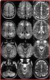
Upper row: 10-year-old, late-diagnosed, intellectually disabled patient with PKU, under treatment (patient number 10). Axial FLAIR (A) and matching DWI (B) and ADC (C) images show increased signal intensity in bilateral parieto-occipital and frontal regions, along with corresponding diffusion restriction. Additionally, a thin linear signal intensity (red arrow) is observed in the splenium of the corpus callosum on D. Lower row: 3-year-old, late-diagnosed, untreated intellectually disabled patient with PKU (patient number 9). Axial FLAIR (E) and matching DWI (F) and ADC (G) images reveal increased signal intensity and restricted diffusion in both globus pallidus. Increased signal intensities near the fourth ventricle are indicated by the red arrows in H. ADC, apparent diffusion coefficient; DWI, diffusion-weighted imaging; FLAIR, fluid-attenuated inversion recovery; PKU, phenylketonuria.
Figure 3. Magnetic resonance imaging of
16-year-old, late-diagnosed, untreated, and intellectually disabled patient with PKU (patient no. 11). Axial FLAIR (A D, G) MRI shows increased signal intensity in the frontal, parietal, occipital, and temporal white matter, also genu and splenium of the corpus callosum (D, red arrows). Matching axial plane DWI (B, E, H) and ADC (C, F, I) maps show restricted diffusion in the corresponding areas. Increased signal intensity on the axial plane FLAIR image (J) is seen in the cerebellum near the fourth ventricle (red arrow). Axial T2W images from the posterior fossa (K) and vertex (L) show slightly enlarged cerebellar folia and cerebral sulci and fissures due to mild cerebellar and cerebral atrophy. ADC, apparent diffusion coefficient; DWI, diffusion-weighted imaging; FLAIR, fluid-attenuated inversion recovery; PKU, phenylketonuria; T2W, T2-weighted.

References

    1. Williams RA, Mamotte CD, Burnett JR. Phenylketonuria: an inborn error of phenylalanine metabolism. Clin Biochem Rev. 2008;29(1):31. - PMC - PubMed
    1. Van Wegberg A, MacDonald A, Ahring K, et al. The complete European guidelines on phenylketonuria: diagnosis and treatment. Orphanet J Rare Dis. 2017;12(1):1 56. - PMC - PubMed
    1. Romani C, Palermo L, MacDonald A, Limback E, Hall SK, Geberhiwot T. The impact of phenylalanine levels on cognitive outcomes in adults with phenylketonuria: effects across tasks and developmental stages. Neuropsychology. 2017;31(3):242 254. (10.1037/neu0000336) - DOI - PMC - PubMed
    1. Hofman DL, Champ CL, Lawton CL, Henderson M, Dye L. A systematic review of cognitive functioning in early treated adults with phenylketonuria. Orphanet J Rare Dis. 2018;13:1 19. - PMC - PubMed
    1. Leuzzi V, Tosetti M, Montanaro D, et al. The pathogenesis of the white matter abnormalities in phenylketonuria. A multimodal 3.0 tesla MRI and magnetic resonance spectroscopy (1 H MRS) study. J Inherit Metab Dis. 2007;30(2):209 216. (10.1007/s10545-006-0399-4) - DOI - PubMed

LinkOut - more resources