. 2023 Sep 4;9(4):00750-2022.
doi: 10.1183/23120541.00750-2022. eCollection 2023 Jul.

Pulmonary artery wedge pressure and left ventricular end-diastolic pressure during exercise in patients with dyspnoea

Affiliations

Pulmonary artery wedge pressure and left ventricular end-diastolic pressure during exercise in patients with dyspnoea

Claudia Baratto et al. ERJ Open Res. .

Abstract

Background: Pulmonary artery wedge pressure (PAWP) during exercise, as a surrogate for left ventricular (LV) end-diastolic pressure (EDP), is used to diagnose heart failure with preserved ejection fraction (HFpEF). However, LVEDP is the gold standard to assess LV filling, end-diastolic PAWP (PAWPED) is supposed to coincide with LVEDP and mean PAWP throughout the cardiac cycle (PAWPM) better reflects the haemodynamic load imposed on the pulmonary circulation. The objective of the present study was to determine precision and accuracy of PAWP estimates for LVEDP during exercise, as well as the rate of agreement between these measures.

Methods: 46 individuals underwent simultaneous right and left heart catheterisation, at rest and during exercise, to confirm/exclude HFpEF. We evaluated: linear regression between LVEDP and PAWP, Bland-Altman graphs, and the rate of concordance of dichotomised LVEDP and PAWP ≥ or < diagnostic thresholds for HFpEF.

Results: At peak exercise, PAWPM and LVEDP, as well as PAWPED and LVEDP, were fairly correlated (R2>0.69, p<0.01), with minimal bias (+2 and 0 mmHg respectively) but large limits of agreement (±11 mmHg). 89% of individuals had concordant PAWP and LVEDP ≥ or <25 mmHg (Cohen's κ=0.64). Individuals with either LVEDP or PAWPM ≥25 mmHg showed a PAWPM increase relative to cardiac output (CO) changes (PAWPM/CO slope) >2 mmHg·L-1·min-1.

Conclusions: During exercise, PAWP is accurate but not precise for the estimation of LVEDP. Despite a good rate of concordance, these two measures might occasionally disagree.


Conflict of interest statement

Conflict of interest: None declared.

Figures
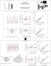
FIGURE 1
FIGURE 1
Accuracy and precision of end-expiratory PAWPED and PAWPM estimates for LVEDP, as well as the rate of agreement of PAWP and LVEDP for the diagnosis of HFpEF at rest and during exercise in our population. The pre-test probability of HFpEF in our cohort was intermediate–high based on the H2FPEF score. Exemplificative pressure trace recordings (LV pressure, red; PAWP, blue) are shown both at rest and during exercise. Linear regression analysis and Bland–Altman plot of end-expiratory LVEDP versus PAWP values are shown both at rest and at peak exercise. Both for rest and peak exercise, a and b) PAWP values are reported both at end-diastole (i.e. mid-A wave for patients in sinus rhythm; mid-C or pre-V wave for patients in atrial fibrillation) and c and d) averaged over the cardiac cycle (mean PAWP). Venn diagrams show the agreement of dichotomised PAWP and LVEDP values above the diagnostic threshold to diagnose HFpEF, both at rest and at peak exercise. Agreement between PAWP and LVEDP was higher during exercise than at rest. Despite substantial agreement at peak exercise, five individuals had either PAWP or LVEDP above the diagnostic threshold for HFpEF. All of them presented with a PAWP/CO slope >2 mmHg·L−1·min−1, suggesting that incorporation of flow-corrected PAWP in the definition of HFpEF (PAWP ≥25 mmHg and/or PAWP/CO slope >2 mmHg·L−1·min−1) may maximise the diagnostic yield of exercise right heart catheterisation. CO: cardiac output; ED: end-diastolic; HFpEF: heart failure with preserved ejection fraction; LV: left ventricular; LVEDP: left ventricular end-diastolic pressure; M: mean; PAWP: pulmonary artery wedge pressure.
FIGURE 2
FIGURE 2
Linear regression analysis and Bland–Altman plot of respiratory-averaged LVEDP versus PAWP values at rest. Bland-Altman plot and linear regression analysis between end-diastolic PAWP (measured at mid-A in sinus rhythm, at mid-C or pre-V in atrial fibrillation) and LVEDP are shown in panels a) and b). Bland–Altman plot and linear regression analysis between mean PAWP (PAWP averaged over the cardiac cycle) and LVEDP are shown in panels c) and d). LVEDP: left ventricular end-diastolic pressure; PAWPED: end-diastolic pulmonary artery wedge pressure; PAWPM: mean pulmonary artery wedge pressure.
FIGURE 3
FIGURE 3
Linear regression analysis and Bland–Altman plot of respiratory-averaged LVEDP versus PAWP values at peak exercise. Bland–Altman plot and linear regression analysis between end-diastolic PAWP (measured at mid-A in sinus rhythm, at mid-C or pre-V in atrial fibrillation) and LVEDP are shown in panels a) and b). Bland–Altman plot and linear regression analysis between mean PAWP (PAWP averaged over the cardiac cycle) and LVEDP are shown in panels c) and d). LVEDP: left ventricular end-diastolic pressure; PAWPED: end-diastolic pulmonary artery wedge pressure; PAWPM: mean pulmonary artery wedge pressure.

References

    1. Borlaug BA, Paulus WJ. Heart failure with preserved ejection fraction: pathophysiology, diagnosis, and treatment. Eur Heart J 2011; 32: 670–679. doi: 10.1093/eurheartj/ehq426 - DOI - PMC - PubMed
    1. Caravita S, Iacovoni A, Senni M. The right side of the circulation in not secondary heart failure with preserved ejection fraction: an elephant in the room? Eur J Heart Fail 2021; 23: 1659–1661. doi: 10.1002/ejhf.2294 - DOI - PubMed
    1. Sorimachi H, Burkhoff D, Verbrugge FH, et al. . Obesity, venous capacitance, and venous compliance in heart failure with preserved ejection fraction. Eur J Heart Fail 2021; 23: 1648–1658. doi: 10.1002/ejhf.2254 - DOI - PubMed
    1. Baratto C, Caravita S, Soranna D, et al. . Current limitations of invasive exercise hemodynamics for the diagnosis of heart failure with preserved ejection fraction. Circ Heart Fail 2021; 14: e007555. doi: 10.1161/CIRCHEARTFAILURE.120.007555 - DOI - PubMed
    1. Borlaug BA, Nishimura RA, Sorajja P, et al. . Exercise hemodynamics enhance diagnosis of early heart failure with preserved ejection fraction. Circ Heart Fail 2010; 3: 588–595. doi:10.1161/CIRCHEARTFAILURE.109.930701 - DOI - PMC - PubMed

LinkOut - more resources