Review
. 2023 Aug 21:10:1245851.
doi: 10.3389/fsurg.2023.1245851. eCollection 2023.

Intra-operative applications of augmented reality in glioma surgery: a systematic review

Affiliations
Review

Intra-operative applications of augmented reality in glioma surgery: a systematic review

Anya Ragnhildstveit et al. Front Surg. .

Abstract

Background: Augmented reality (AR) is increasingly being explored in neurosurgical practice. By visualizing patient-specific, three-dimensional (3D) models in real time, surgeons can improve their spatial understanding of complex anatomy and pathology, thereby optimizing intra-operative navigation, localization, and resection. Here, we aimed to capture applications of AR in glioma surgery, their current status and future potential.

Methods: A systematic review of the literature was conducted. This adhered to the Preferred Reporting Items for Systematic Reviews and Meta-Analyses (PRISMA) guideline. PubMed, Embase, and Scopus electronic databases were queried from inception to October 10, 2022. Leveraging the Population, Intervention, Comparison, Outcomes, and Study design (PICOS) framework, study eligibility was evaluated in the qualitative synthesis. Data regarding AR workflow, surgical application, and associated outcomes were then extracted. The quality of evidence was additionally examined, using hierarchical classes of evidence in neurosurgery.

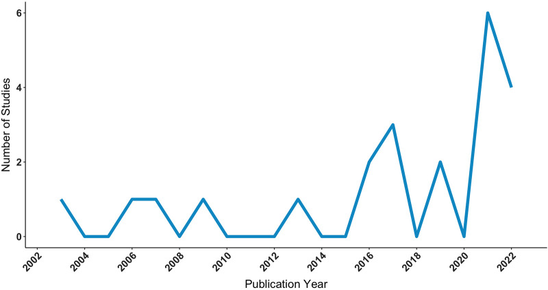
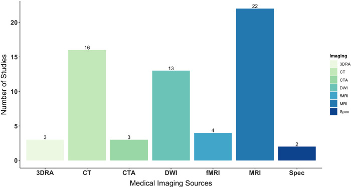
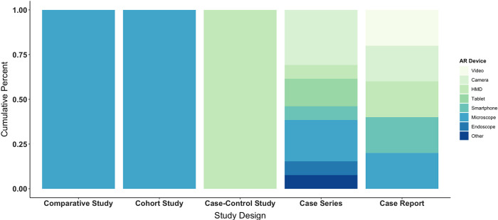
Results: The search returned 77 articles. Forty were subject to title and abstract screening, while 25 proceeded to full text screening. Of these, 22 articles met eligibility criteria and were included in the final review. During abstraction, studies were classified as "development" or "intervention" based on primary aims. Overall, AR was qualitatively advantageous, due to enhanced visualization of gliomas and critical structures, frequently aiding in maximal safe resection. Non-rigid applications were also useful in disclosing and compensating for intra-operative brain shift. Irrespective, there was high variance in registration methods and measurements, which considerably impacted projection accuracy. Most studies were of low-level evidence, yielding heterogeneous results.

Conclusions: AR has increasing potential for glioma surgery, with capacity to positively influence the onco-functional balance. However, technical and design limitations are readily apparent. The field must consider the importance of consistency and replicability, as well as the level of evidence, to effectively converge on standard approaches that maximize patient benefit.

Keywords: augmented reality; brain tumor; glioma; mixed reality; neuronavigation; neurosurgery; systematic review; virtual reality.


Conflict of interest statement

The authors declare that the research was conducted in the absence of any commercial or financial relationships that could be construed as a potential conflict of interest.

Figures

Figure 1
Figure 1
Number of studies applying AR in glioma surgery by publication year, as identified via PubMed, Embase, and Scopus electronic database searches, executed on October 22, 2022. AR, augmented reality.
Figure 2
Figure 2
PRISMA 2020 flow diagram, describing the search strategy and selection schema of the review process.
Figure 3
Figure 3
Number of patients diagnosed with glioma recruited in AR studies by study type, excluding phantom patients (A). Medical image acquisition phase by study type, spanning pre-operative and/or intra-operative stages (B). Study types are grouped by primary aim into “development” or “intervention”, with respect to AR application. AR, augmented reality.
Figure 4
Figure 4
Types of medical imaging used as data sources for AR, specifically segmenting, modeling, formatting, and projecting virtual objects onto phantoms or patients’ real anatomy. AR, augmented reality.
Figure 5
Figure 5
Types of augmented reality display devices used across study designs, including comparative, cohort, and case-control studies as well as case series and case reports. AR, augmented reality; HMD, head-mounted display.
Figure 6
Figure 6
Illustrative case. Primary motor cortex glioblastoma of the dominant hemisphere. Intraoperative photographs obtained under white light (A), YELLOW 560 filter (B), and AR HDFT (C) before tumor resection. (D–I) The main steps of the surgery that were performed in large part along with the AR HDFT-F technique. (J–L) The surgical field at the end of the tumor resection obtained under white light (J), combined INFRARED 800 and AR HDFT during indocyanine green videoangiography (K), and AR HDFT-F (L). Insets in panels (C,F,I,L) are the screenshots obtained during the microscope focus-based neuronavigation. “Supratentorial High-Grade Gliomas: Maximal Safe Anatomical Resection Guided by Augmented Reality High-Definition Fiber Tractography and Fluorescein.” This figure is protected by Copyright, is owned by The Journal of Neurosurgery Publishing Group (JNSPG), and is used with permission only within this document. Permission to use it otherwise must be secured from JNSPG. Full text of the article containing the original figure is available at thejns.org.
Figure 7
Figure 7
Illustrative case. A 31-year-old man with oligodendroglioma. (A) Intraoperative brain surface photograph in JPEG (Joint Photographic Experts Group) format. (B) Fusion 3-dimensional computer graphics (3DCG) created from preoperative imaging studies. The purple highlight indicates the tumor area. (C) Mixed-reality computer graphics created by aligning the intraoperative brain surface photograph and fusion 3DCG. The purple highlight indicates the tumor area. “Development of Innovative Neurosurgical Operation Support Method Using Mixed-Reality Computer Graphics.” © 2021 The Author(s). Published by Elsevier Inc. Licensed under CC-BY-NC-ND.

References

    1. American Association of Neurological Surgeon. Brain Tumors. Available at: https://www.aans.org/en/Patients/Neurosurgical-Conditions-and-Treatments... (Accessed August 19, 2021).
    1. Ostrom QT, Bauchet L, Davis FG, Deltour I, Fisher JL, Langer CE, et al. The epidemiology of glioma in adults: a “state of the science” review. Neuro-Oncology. (2014) 16(7):896–913. 10.1093/neuonc/nou087 - DOI - PMC - PubMed
    1. Lin D, Wang M, Chen Y, Gong J, Chen L, Shi X, et al. Trends in intracranial glioma incidence and mortality in the United States, 1975–2018. Front Oncol. (2021) 11:748061. 10.3389/fonc.2021.748061 - DOI - PMC - PubMed
    1. Gerritsen JKW, Broekman MLD, De Vleeschouwer S, Schucht P, Nahed BV, Berger MS, et al. Safe surgery for glioblastoma: recent advances and modern challenges. Neurooncol Pract. (2022) 9(5):364–79. 10.1093/nop/npac019 - DOI - PMC - PubMed
    1. Rabab’h O, Al-Ramadan A, Shah J, Lopez-Negrete H, Gharaibeh A. Twenty years after glioblastoma multiforme diagnosis: a case of long-term survival. Cureus. (2021) 13(6). 10.7759/cureus.16061 - DOI - PMC - PubMed