

Vaccination with the Crimean-Congo hemorrhagic fever virus viral replicon vaccine induces NP-based T-cell activation and antibodies possessing Fc-mediated effector functions
- PMID: 37671145
- PMCID: PMC10475602
- DOI: 10.3389/fcimb.2023.1233148
Vaccination with the Crimean-Congo hemorrhagic fever virus viral replicon vaccine induces NP-based T-cell activation and antibodies possessing Fc-mediated effector functions
Abstract
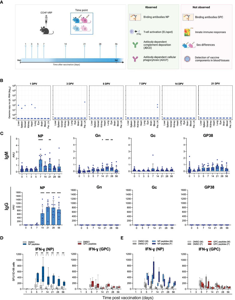
Crimean-Congo hemorrhagic fever virus (CCHFV; family Nairoviridae) is a tick-borne pathogen that frequently causes lethal disease in humans. CCHFV has a wide geographic distribution, and cases have been reported in Africa, Asia, the Middle East, and Europe. Availability of a safe and efficacious vaccine is critical for restricting outbreaks and preventing disease in endemic countries. We previously developed a virus-like replicon particle (VRP) vaccine that provides complete protection against homologous and heterologous lethal CCHFV challenge in mice after a single dose. However, the immune responses induced by this vaccine are not well characterized, and correlates of protection remain unknown. Here we comprehensively characterized the kinetics of cell-mediated and humoral immune responses in VRP-vaccinated mice, and demonstrate that they predominantly target the nucleoprotein (NP). NP antibodies are not associated with protection through neutralizing activity, but VRP vaccination results in NP antibodies possessing Fc-mediated antibody effector functions, such as complement activation (ADCD) and antibody-mediated cellular phagocytosis (ADCP). This suggests that Fc-mediated effector functions may contribute to this vaccine's efficacy.
Keywords: CCHFV; Fc-mediated effector function; antibody; nucleoprotein; vaccine.
Copyright © 2023 Scholte, Karaaslan, O’Neal, Sorvillo, Genzer, Welch, Coleman-McCray, Spengler, Kainulainen, Montgomery, Pegan, Bergeron and Spiropoulou.
Conflict of interest statement
Provisional US Patent Application No. 62/780,098. The authors declare that the research was conducted in the absence of any commercial or financial relationships that could be construed as a potential conflict of interest.
Figures
Similar articles
-
Replicon particle vaccination induces non-neutralizing anti-nucleoprotein antibody-mediated control of Crimean-Congo hemorrhagic fever virus.NPJ Vaccines. 2024 May 23;9(1):88. doi: 10.1038/s41541-024-00877-1. NPJ Vaccines. 2024. PMID: 38782933 Free PMC article.
-
Nucleoside-Modified mRNA Vaccines Protect IFNAR-/- Mice against Crimean-Congo Hemorrhagic Fever Virus Infection.J Virol. 2022 Feb 9;96(3):e0156821. doi: 10.1128/JVI.01568-21. Epub 2021 Nov 24. J Virol. 2022. PMID: 34817199 Free PMC article.
-
Crimean-Congo hemorrhagic fever virus replicon particle vaccine is safe and elicits functional, non-neutralizing anti-nucleoprotein antibodies and T cell activation in rhesus macaques.Antiviral Res. 2025 Jan;233:106045. doi: 10.1016/j.antiviral.2024.106045. Epub 2024 Dec 1. Antiviral Res. 2025. PMID: 39626793
-
Recent advances in understanding Crimean-Congo hemorrhagic fever virus.F1000Res. 2018 Oct 29;7:F1000 Faculty Rev-1715. doi: 10.12688/f1000research.16189.1. eCollection 2018. F1000Res. 2018. PMID: 30416710 Free PMC article. Review.
-
Crimean-Congo Hemorrhagic Fever Virus: Progress in Vaccine Development.Diagnostics (Basel). 2023 Aug 19;13(16):2708. doi: 10.3390/diagnostics13162708. Diagnostics (Basel). 2023. PMID: 37627967 Free PMC article. Review.
Cited by
-
Rapid, sensitive, and species-independent detection of Crimean Congo hemorrhagic fever virus nucleoprotein and GP38 antibodies.EBioMedicine. 2025 Aug;118:105857. doi: 10.1016/j.ebiom.2025.105857. Epub 2025 Jul 23. EBioMedicine. 2025. PMID: 40706445 Free PMC article.
-
Replicon particle vaccination induces non-neutralizing anti-nucleoprotein antibody-mediated control of Crimean-Congo hemorrhagic fever virus.NPJ Vaccines. 2024 May 23;9(1):88. doi: 10.1038/s41541-024-00877-1. NPJ Vaccines. 2024. PMID: 38782933 Free PMC article.
-
Cellular immunity to nucleoproteins (NP) of Crimean-Congo hemorrhagic fever virus (CCHFV) and Hazara Virus (HAZV).Med Microbiol Immunol. 2024 Sep 25;213(1):20. doi: 10.1007/s00430-024-00802-2. Med Microbiol Immunol. 2024. PMID: 39320473
-
Prime-Boost Vaccination Based on Nanospheres and MVA Encoding the Nucleoprotein of Crimean-Congo Hemorrhagic Fever Virus Elicits Broad Immune Responses.Vaccines (Basel). 2025 Mar 10;13(3):291. doi: 10.3390/vaccines13030291. Vaccines (Basel). 2025. PMID: 40266214 Free PMC article.
-
Reverse Genetics System for Crimean-Congo Hemorrhagic Fever Virus.Methods Mol Biol. 2025;2893:247-256. doi: 10.1007/978-1-0716-4338-9_18. Methods Mol Biol. 2025. PMID: 39671042
References
Publication types
MeSH terms
Substances
Grants and funding
LinkOut - more resources
Full Text Sources
Medical
Miscellaneous