

Inadvertent catheter misplacement into the subclavian artery during ultrasound-guided internal jugular venous catheterization: a case report
- PMID: 37672125
- PMCID: PMC10482804
- DOI: 10.1186/s40981-023-00649-1
Inadvertent catheter misplacement into the subclavian artery during ultrasound-guided internal jugular venous catheterization: a case report
Abstract
Background: Ultrasound-guided central venous catheterization has become a standard procedure. However, mechanical complications are still reported.
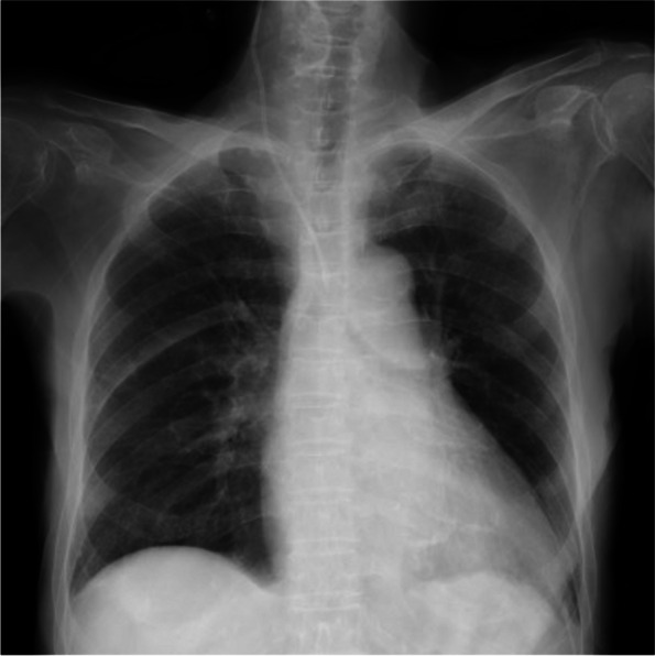
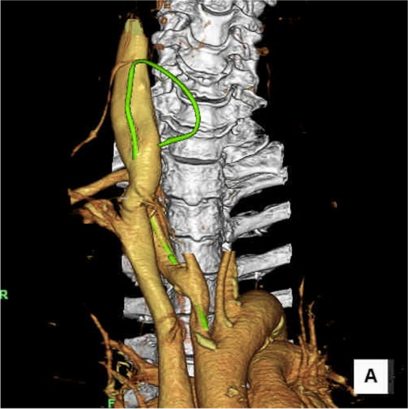
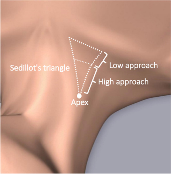
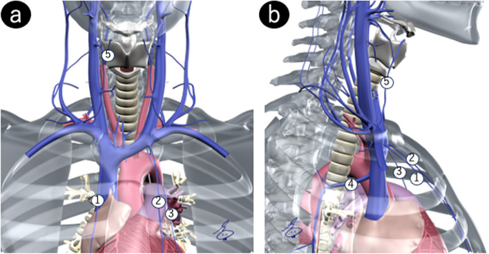
Case presentation: An 85-year-old woman presented with coagulopathic bladder tamponade. Ultrasound-guided right internal jugular venous catheterization was planned because of difficult peripheral venous access. A guidewire was advanced through a needle inserted at the midpoint of the right carotid triangle. The guidewire was identified in the short axis, but not in the long-axis ultrasound view, leading to inadvertent insertion of the catheter into the right subclavian artery through the internal jugular vein. Stent graft insertion was performed for perforation closure. The patient exhibited no symptoms of cerebral ischemia following stent graft insertion.
Discussion: This case demonstrated that the needle-sticking site should not be placed close to the clavicle for ultrasound-guided internal jugular venous catheterization, as it may not confirm the position of guidewire in the long-axis ultrasound view.
Keywords: Guidewire; Low approach; Mechanical complication; Ultrasound-guided internal jugular venous catheterization.
© 2023. The Japanese Society of Anesthesiologists.
Conflict of interest statement
J. T. is a technical adviser of the Cardinal Health K.K. (Japan) and has done an ultrasound-guided technical training course held by the company. The other authors declare that they have no competing interests.
Figures
References
-
- Lee YH, Kim TK, Jung YS, Cho YJ, Yoon S, Seo JH, et al. Comparison of needle insertion and guidewire placement techniques during internal jugular vein catheterization: the thin-wall introducer needle technique versus the cannula-over-needle technique. Crit Care Med. 2015;43(10):2112–2116. doi: 10.1097/CCM.0000000000001167. - DOI - PubMed
LinkOut - more resources
Full Text Sources