. 2023 Nov 6;222(11):e200212128072023new.
doi: 10.1083/jcb.200212128072023new. Epub 2023 Sep 6.

The RasGAP-associated endoribonuclease G3BP mediates stress granule assembly

Affiliations

The RasGAP-associated endoribonuclease G3BP mediates stress granule assembly

Hélène Tourrière et al. J Cell Biol. .

Abstract

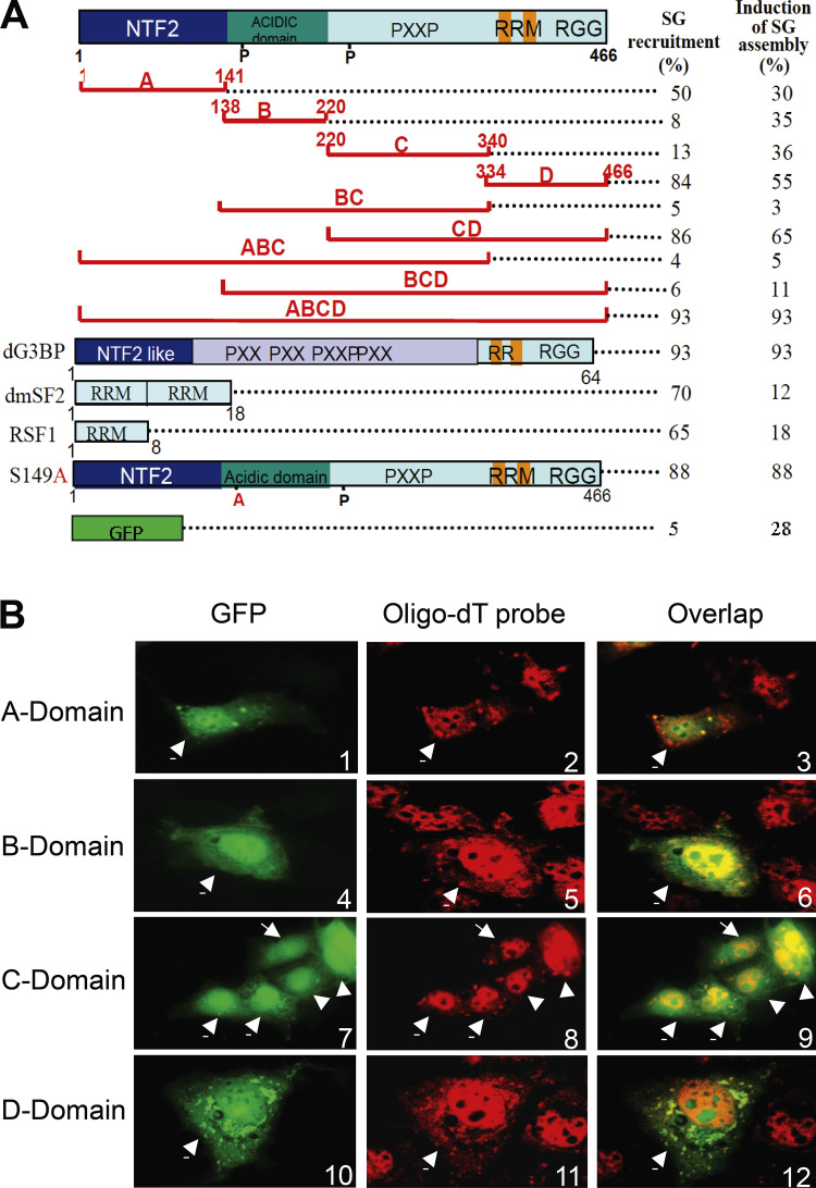
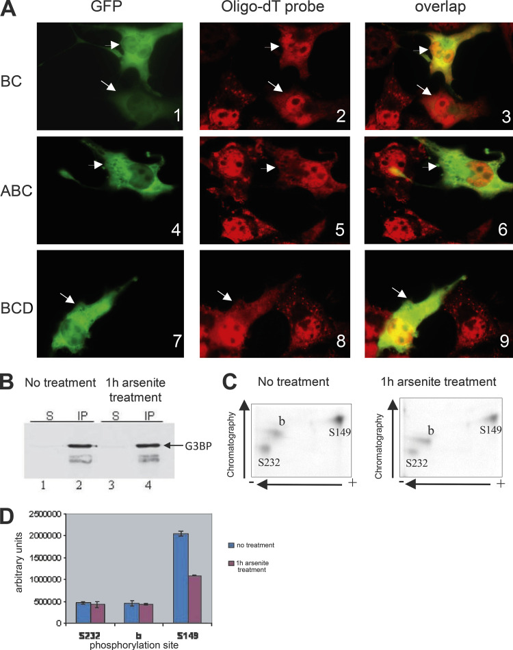
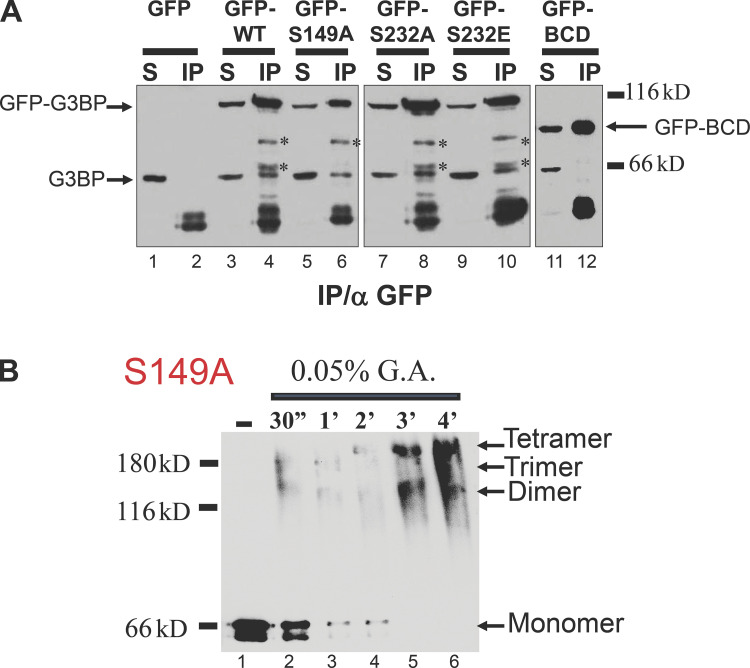
Stress granules (SGs) are formed in the cytoplasm in response to various toxic agents and are believed to play a critical role in the regulation of mRNA metabolism during stress. In SGs, mRNAs are stored in an abortive translation initiation complex that can be routed to either translation initiation or degradation. Here, we show that G3BP, a phosphorylation-dependent endoribonuclease that interacts with RasGAP, is recruited to SGs in cells exposed to arsenite. G3BP may thus determine the fate of mRNAs during cellular stress. Remarkably, SG assembly can be either dominantly induced by G3BP overexpression, or on the contrary, inhibited by expressing a central domain of G3BP. This region binds RasGAP and contains serine 149 whose dephosphorylation is induced by arsenite treatment. Critically, a non-phosphorylatable G3BP mutant (S149A) oligomerizes and assembles SG. These results suggest that G3BP is an effector of SG assembly and that Ras signaling contributes to this process by regulating G3BP dephosphorylation.


Conflict of interest statement

Disclosures: The authors declare no competing interests exist.

Figures

Figure 1.
Figure 1.
G3BP is recruited to SGs. (A) Fixed Cos cells were stained with an anti-G3BP antibody (1 and 2) or transfected with Drosophila G3BP-GFP (dG3BP-GFP, 3) fusion (green) and a fluorescent oligo-dT probe to reveal SGs (red). (B) Intracellular localization of G3BP-GFP fusion (green) in transfected HeLa cells treated with 0.5 mM arsenite for 1 h, which were fixed and stained with anti-TIA1/R antibody (red). Arrows indicate transfected cells expressing G3BP-GFP fusion. (C) Fixed CCL39 and Ha-Ras cells were treated with 1 mM arsenite for 20 min and stained with an anti-G3BP antibody (green) and a fluorescent oligo-dT probe to reveal SGs (red).
Figure 2.
Figure 2.
G3BP domains A and D can direct GFP fusion proteins to arsenite-induced SGs. (A) Efficiency of recruitment to SGs. Schematic representation of the GFP fusion proteins; G3BP domain A, B, C, and D, and the phosphorylation mutant S149A (see text); G3BP from Drosophila (dG3BP); RRM of dmSF2 and RSF1. Numbers refer to the first and the last residue of each region. Transfected Cos cells were scored (100 transfected cells averaged from two experiments) for the ability of each GFP fusion to either be recruited to SGs after arsenite treatment (left column) or dominantly induce SGs assembly without treatment (right column). In each case, hybridization with oligo-dT probe was included to positively identify SGs. (B) Intracellular localization of G3BP domains fused to GFP (green). SGs were visualized with fluorescent oligo-dT probes (red). Arrows indicate transfected cells expressing GFP fusions.
Figure 3.
Figure 3.
G3BP BC domain inhibits arsenite-induced assembly of SGs. (A) Cos cells were transiently transfected with GFP-BC, GFP-ABC, or GFP-BCD, treated with arsenite before fixation, and double stained with fluorescent oligo-dT probe (red). GFP, green. Arrows indicate transfected cells expressing GFP fusion. (B) Equal amounts of whole-cell extracts prepared from metabolically labeled CCL39 cells were immunoprecipitated with anti-G3BP antibody. Proteins were revealed by immunoblot analysis using the same antibody. S, supernatant; IP, immunoprecipitation. (C) Phosphotryptic peptide mapping of immunopurified 32P-labeled G3BP, shown in B, from untreated (left) and arsenite-treated (right) CCL39 cells. The identification of phosphorylation sites Ser 149 and Ser 232 was previously reported (Tourrière et al., 2001). (D) The intensity of each spot from untreated (blue) and arsenite-treated (red) was quantitated by densitometry scanning of the chromatography plates using ImageQuant software version 5.2. Error bars resulting from two independently performed experiments are shown.
Figure 4.
Figure 4.
G3BP assembles SGs. Untreated Cos cells transfected with GFP or G3BP-GFP (green) were counterstained with either fluorescent oligo-dT (2 and 5, red) or anti-HuR antibody (8, red). Untreated HeLa cells transfected with G3BP-GFP (10, green) were counterstained with anti-TIA1/R antibody (11, red).
Figure 5.
Figure 5.
G3BP oligomerization in vitro. (A) Whole-cell extracts prepared from Cos cells transfected with GFP, GFP-G3BP wild type, phosphorylation mutants S149A, S232A, S232E, and BCD-mutant lacking A domain were immunoprecipitated with anti-GFP antibodies. Proteins were revealed by immunoblot analysis using anti-G3BP antibody. S, supernatant; IP, immunoprecipitation. Asterisks correspond to proteolytic fragments of G3BP-GFP that can be detected by anti-GFP antibodies, whereas the band at the level of G3BP is only seen with the antiG3BP antibody. (B) Glutaraldehyde crosslinking analysis of purified G3BP phosphorylation mutants S149A. Proteins (0.2 g each) were incubated for the indicated time with glutaraldehyde (G.A.) and were then analyzed by Western blotting with an anti-G3BP.

Corrected and republished from

  • doi: 10.1083/jcb.200212128

References

    1. Allemand, E., Gattoni R., Bourbon H.M., Stevenin J., Cáceres J.F., Soret J., and Tazi J.. 2001. Distinctive features of Drosophila alternative splicing factor RS domain: Implication for specific phosphorylation, shuttling, and splicing activation. Mol. Cell. Biol. 21:1345–1359. 10.1128/MCB.21.4.1345-1359.2001 - DOI - PMC - PubMed
    1. Allemand, E., Dokudovskaya S., Bordonné R., and Tazi J.. 2002. A conserved Drosophila transportin-serine/arginine-rich (SR) protein permits nuclear import of Drosophila SR protein splicing factors and their antagonist repressor splicing factor 1. Mol. Biol. Cell. 13:2436–2447. 10.1091/mbc.e02-02-0102 - DOI - PMC - PubMed
    1. Bullock, T.L., Clarkson W.D., Kent H.M., and Stewart M.. 1996. The 1.6 angstroms resolution crystal structure of nuclear transport factor 2 (NTF2). J. Mol. Biol. 260:422–431. 10.1006/jmbi.1996.0411 - DOI - PubMed
    1. Gallouzi, I.E., Parker F., Chebli K., Maurier F., Labourier E., Barlat I., Capony J.P., Tocque B., and Tazi J.. 1998. A novel phosphorylation-dependent RNase activity of GAP-SH3 binding protein: A potential link between signal transduction and RNA stability. Mol. Cell. Biol. 18:3956–3965. 10.1128/MCB.18.7.3956 - DOI - PMC - PubMed
    1. Kedersha, N.L., Gupta M., Li W., Miller I., and Anderson P.. 1999. RNA-binding proteins TIA-1 and TIAR link the phosphorylation of eIF-2 alpha to the assembly of mammalian stress granules. J. Cell Biol. 147:1431–1442. 10.1083/jcb.147.7.1431 - DOI - PMC - PubMed

Publication types

MeSH terms