. 2023 Dec;193(12):2182-2202.
doi: 10.1016/j.ajpath.2023.08.004. Epub 2023 Sep 4.

The Sympathetic Nervous System Promotes Hepatic Lymphangiogenesis, which Is Protective Against Liver Fibrosis

Affiliations

The Sympathetic Nervous System Promotes Hepatic Lymphangiogenesis, which Is Protective Against Liver Fibrosis

Masatake Tanaka et al. Am J Pathol. 2023 Dec.

Abstract

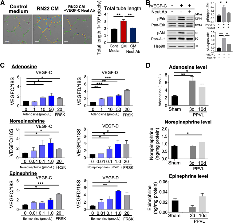
Liver is the largest lymph-producing organ. In cirrhotic patients, lymph production significantly increases concomitant with lymphangiogenesis. The aim of this study was to determine the mechanism of lymphangiogenesis in liver and its implication in liver fibrosis. Liver biopsies from portal hypertensive patients with portal-sinusoidal vascular disease (n = 22) and liver cirrhosis (n = 5) were evaluated for lymphangiogenesis and compared with controls (n = 9 and n = 6, respectively). For mechanistic studies, rats with partial portal vein ligation (PPVL) and bile duct ligation (BDL) were used. A gene profile data set (GSE77627), including 14 histologically normal liver, 18 idiopathic noncirrhotic portal hypertension, and 22 cirrhotic patients, was analyzed. Lymphangiogenesis was significantly increased in livers from patients with portal-sinusoidal vascular disease, cirrhotic patients, as well as PPVL and BDL rats. Importantly, Schwann cells of sympathetic nerves highly expressed vascular endothelial growth factor-C in PPVL rats. Vascular endothelial growth factor-C neutralizing antibody or sympathetic denervation significantly decreased lymphangiogenesis in livers of PPVL and BDL rats, which resulted in progression of liver fibrosis. Liver specimens from cirrhotic patients showed a positive correlation between sympathetic nerve/Schwann cell-positive areas and lymphatic vessel numbers, which was supported by gene set analysis from patients with noncirrhotic portal hypertension and cirrhotic patients. Sympathetic nerves promote hepatic lymphangiogenesis in noncirrhotic and cirrhotic livers. Increased hepatic lymphangiogenesis can be protective against liver fibrosis.


Figures

None
Graphical abstract
Figure 1
Figure 1
Hepatic lymphangiogenesis occurs in livers from patients with portal-sinusoidal vascular disease (PSVD) and rats with partial portal vein ligation (PPVL). A: Left panels: Podoplanin (PDPN; a marker of lymphatic vessels) immunohistochemistry images in controls and patients with noncirrhotic portal hypertension resulting from presinusoidal vascular disease (PSVD). Right panel: Lymphatic vessel (LV) number was quantified in controls and patients with PSVD. B: Immunofluorescence (IF) images of lymphatic vessel endothelial hyaluronan receptor 1 (LYVE-1; a marker of LVs; red), α-smooth muscle actin (α-SMA; a marker of blood vessels; green), and DAPI (a marker of nuclei; blue) in sham and 10-day PPVL rat livers. Arrows indicate lymphatic vessels. C: Quantification of LV number (left panel) and area (right panel) in sham and PPVL rat livers. D: Evaluation of proliferating LVs as an indicator of lymphangiogenesis. IF images of proliferating cell nuclear antigen (PCNA; green) and prospero homeobox 1 (Prox-1; another marker of LVs; red; left panel) or LYVE-1 (right panel) in 3-day PPVL rat livers. Double-positive cells (arrows) are proliferating lymphatic endothelial cells (LyECs). E: Quantification of PCNA-positive LyECs in sham and PPVL rat livers. n = 9 controls (A); n = 22 patients with PSVD (A); n = 5 per group (C and E). ∗P < 0.05, ∗∗P < 0.01, and ∗∗∗P < 0.001. Scale bars: 100 μm (A); 20 μm (B and D). BD, bile duct; HA, hepatic arteriole; PV, portal venule.
Figure 2
Figure 2
Schwann cells of sympathetic nerves are the major source of vascular endothelial growth factor (VEGF)-C, promoting hepatic lymphangiogenesis in partial portal vein ligation (PPVL) rats. A: Immunofluorescence (IF) images of VEGF-C (red) and DAPI (blue) in sham or 3- and 10-day PPVL rat livers. Arrows indicate VEGF-C–positive cell clusters. B: VEGF-C level in sham and PPVL rat whole liver homogenates. C: Top panels: IF images of lymphatic vessel endothelial hyaluronan receptor 1 (LYVE-1; red) and DAPI (blue) in 10-day PPVL rat livers with treatment of control rabbit IgG or VEGF-C–neutralizing antibody (Neut Ab). Arrows indicate lymphatic vessels (LVs). Bottom panels: VEGF-C–neutralizing antibody treatment significantly decreased lymphangiogenesis in 10-day PPVL rat livers. D: IF images of tyrosine hydroxylase (TH; a marker of sympathetic nerves; green; D1), S100 calcium binding protein (S-100; a marker of Schwann cells; green; D2), acetylcholine esterase (AChE; a marker of parasympathetic nerves; green; D3), VEGF-C (red), and DAPI (blue) in 3-day PPVL rat livers. Colocalization of VEGF-C with TH, S-100, and AChE was confirmed by line profile analysis. Fluorescence intensity of line profile graphs (right panels) correspond to the white arrows drawn on enlarged images (left panels). Yellow arrows indicate VEGF-C colocalization cluster with TH or S-100. n = 5 per group (B); n = 6 per group (C). ∗P < 0.05, ∗∗P < 0.01, and ∗∗∗P < 0.001. Scale bars = 20 μm (A, C, and D). HA, hepatic arteriole; PV, portal venule.
Figure 3
Figure 3
Rat Schwann cells induce lymphangiogenesis by producing prolymphangiogenic factors, such as vascular endothelial growth factor (VEGF)-C and VEGF-D. A: Human dermal lymphatic endothelial cells (hDLECs) incubated with control medium and RN22 rat Schwann cell conditioned medium (CM) with or without 20 μg/mL VEGF-C–neutralizing antibody (Neut Ab) for 4 hours. Left panels: Five images were taken per well and analyzed by the angiogenesis analyzer macro of ImageJ software version 1.53o (NIH, Bethesda, MD). Right panel: Quantification of the total length of cellular cords. Representative data from three independent experiments are shown. B: Validation of VEGF-C–neutralizing antibody. hDLEC was incubated with human recombinant VEGF-C (final concentration, 50 ng/mL) or VEGF-C–neutralizing antibody (50 μg/mL) for 30 minutes. Left panel: Western blot analysis images of phosphorylated extracellular signal-regulated kinase (pErk) and phosphorylated Akt (pAkt) in hDLEC treated with human recombinant VEGF-C or VEGF-C–neutralizing antibody. Right panels: Quantitations of pErk and pAkt protein levels normalized with pan-Erk and pan-Akt are shown, respectively. C: VEGF-C and VEGF-D expression in rat Schwann cells (RN22) treated with adenosine or catecholamines (norepinephrine and epinephrine) at indicated concentrations for 4 hours. Forskolin (FRSK; 20 μmol/L), an adenylyl cyclase activator, was used as a positive control. Representative data from three independent experiments are shown. D: Adenosine, norepinephrine, and epinephrine levels in sham and partial portal vein ligation (PPVL) rat livers. n = 3 (AC); n = 7 for sham and 10-day PPVL (D); n = 5 for 3-day PPVL (D). ∗P < 0.05, ∗∗P < 0.01, and ∗∗∗P < 0.001. Scale bars = 100 μm (A). Cont, control; Hsp90, heat shock protein 90.
Figure 4
Figure 4
Sympathetic nerve/Schwann cell–positive areas are positively related to the number of lymphatic vessels in the livers of partial portal vein ligation (PPVL) rats. A: Immunofluorescence images of tyrosine hydroxylase (TH; a marker of sympathetic nerves; red), ubiquitin C-terminal hydrolase L1 (PGP9.5; a marker of panneurons; green), and DAPI (blue) in sham and PPVL rat livers. B: Quantification of TH-positive areas in sham and PPVL rat livers. There was a trend of decreased TH+ area 30 days after PPVL compared with that of 10 days after PPVL (P = 0.06). C: A correlation between sympathetic nerve–positive areas (TH+) and lymphatic vessel (LV) numbers. D: Quantification of S100 calcium binding protein B (S100B; a marker of Schwann cells)–positive areas in sham and PPVL rat livers. E: A correlation between Schwann cell–positive areas (TH+/S100B+) and LV number. F: Three-dimensional images of sympathetic nerves (TH; red) and LVs [lymphatic vessel endothelial hyaluronan receptor 1 (LYVE-1); white] in sham and 10-day PPVL rat livers. Insets: Indicated LV is enlarged. n = 5 for sham and 3-, 10-, and 30-day PPVL (B and D); n = 4 for 1-day PPVL (B and D); n = 20 (C and E); n = 1 per group (F). ∗P < 0.05, ∗∗P < 0.01. Scale bars = 100 μm (A and F). HA, hepatic arteriole; PV, portal venule.
Figure 5
Figure 5
Inhibition of sympathetic nerve activity suppresses lymphangiogenesis in the livers of partial portal vein ligation (PPVL) rats. A: Macroscopic image of the celiac ganglion. The celiac ganglion (arrowheads in a and b) was identified between the aorta and the celiac artery (CA) by toluidine blue (TB) topical application (b), TB stain (c), and tyrosine hydroxylase (TH) immunofluorescence (d). B: Schematic diagram showing celiac ganglionectomy (CG). C: Left panels: Immunofluorescence (IF) images of lymphatic vessel endothelial hyaluronan receptor 1 (LYVE-1; red) and DAPI (blue) in 3- and 10-day PPVL rat livers with or without CG. Arrows indicate lymphatic vessels (LVs). Right panels: CG significantly decreased VEGF-C level and lymphangiogenesis in 3- and 10-day PPVL rat livers. D: Left panels: IF images of LYVE-1 (red) and DAPI (blue) in 3- and 10-day PPVL rat liver with or without 6-hydroxydopamine (6-OHDA) administration. Yellow arrows indicate lymphatic vessels. Right panels: 6-OHDA administration significantly decreased VEGF-C levels and lymphangiogenesis in 3- and 10-day PPVL rat livers. n = 5 per group (C and D). ∗P < 0.05, ∗∗P < 0.01, and ∗∗∗P < 0.001. Scale bars: 200 μm (A); 100 μm (C and D). BD, bile duct; Cont, control; HA, hepatic arteriole; PV, portal venule.
Figure 6
Figure 6
Macrophages play a role in hepatic lymphangiogenesis in partial portal vein ligation (PPVL) rats independently of vascular endothelial growth factor (VEGF)-C synthesis. A: Immunofluorescence (IF) images of CD68-positive macrophages (red), α-smooth muscle actin (α-SMA; green), and DAPI (blue) in sham and 10-day PPVL rat livers. B: Quantification of CD68-positive macrophages in sham and PPVL rat livers. C: Left panels: IF images of CD68-positive macrophages (red) and DAPI (blue) in 3- and 10-day PPVL rat liver with or without celiac ganglionectomy (CG). Right panels: CG significantly decreased CD68-positive macrophage number in 3- and 10-day PPVL rat livers. D: Left panels: IF images of CD68-positive macrophages (red) and DAPI (blue) in 3- and 10-day PPVL rat liver with or without 6-hydroxydopamine (6-OHDA) administration. Right panels: 6-OHDA administration significantly decreased CD68-positive macrophage number in 3- and 10-day PPVL rat livers. E: Top panels: IF images of lymphatic vessel endothelial hyaluronan receptor 1 (LYVE-1; red), CD68 (green), and DAPI (blue) in 10-day PPVL rat livers with or without clodronate-liposome (Clod-Lp) administration. Arrows indicate lymphatic vessels. Bottom panels: Clod-Lp administration significantly decreased lymphangiogenesis in 10-day PPVL rat livers. F: IF images of VEGF-C (red), CD68 (green), and DAPI (blue) in sham or 3- and 10-day PPVL rat livers. G: Flow cytometry of nonparenchymal cell fractions isolated from sham or 3- and 10-day PPVL rat livers. Double-positive cells for CD68 and VEGF-C were not significantly observed in either sham or 3- and 10-day PPVL rat livers. Representative data of three independent experiments are shown. n = 5 per group (BE). ∗P < 0.05, ∗∗P < 0.01, and ∗∗∗P < 0.001. Scale bars: 20 μm (A and E); 100 μm (C and D); 50 μm (F). Cont, control; PV, portal venule.
Figure 7
Figure 7
Lymphatic vessels are significantly increased and positively related to Schwann cell abundance in human cirrhotic livers. A: Left panels: Podoplanin (PDPN; a marker of lymphatic vessels) immunohistochemistry images in controls and patients with cirrhotic portal hypertension (CPH) resulting from alcohol-associated cirrhosis. Arrows indicate lymphatic vessels (LVs). Right panel: LV number was quantified in controls and patients with CPH. B: Left panels: Immunofluorescence images of tyrosine hydroxylase (TH; a marker of sympathetic nerves; red) and S100b (a marker of Schwann cells; green) in controls and patients with CPH. Right panel: Schwann cells (TH+/S100b+) of sympathetic nerves were quantified in controls and patients with CPH. C: A correlation between Schwann cells and LV number in human livers. n = 6 controls (A and B); n = 5 patients with CPH (A and B); n = 10 (C). ∗P < 0.05. ∗∗P < 0.01. Scale bars: 100 μm (A); 20 μm (B). PV, portal venule.
Figure 8
Figure 8
Denervation of sympathetic nerves significantly decreases hepatic lymphatic vessel (LV) numbers and increases liver fibrosis in rats with bile duct ligation (BDL). A: Immunofluorescence (IF) images of tyrosine hydroxylase (TH; red) and DAPI (blue) in sham, celiac ganglionectomy (CG), and BDL rat livers with or without CG (left panels) as well as their quantification (right panel). B: IF images of lymphatic vessel endothelial hyaluronan receptor 1 (LYVE-1; red) and DAPI (blue) in sham, CG, and BDL rat livers with or without CG (left panels) as well as their quantification (right panel). Arrows indicate lymphatic vessels. C: A correlation between TH-positive area and LV area. D: Evaluation of liver fibrosis. Sirius red staining (left panels) and its quantification (right top panel) as well as hydroxyproline measurement (right bottom panel). E: Schematic illustration of the potential mechanism of lymphangiogenesis in liver. n = 7 sham, BDL, and BDL + CG (AD); n = 6 CG (A and B); n = 25 (C); n = 7 CG (D). ∗P < 0.05, ∗∗P < 0.01, and ∗∗∗P < 0.001. Scale bars = 100 μm (A, B, and D). BD, bile ductule; HA, hepatic arteriole; PV, portal venule.
Figure 9
Figure 9
Sympathetic nerve marker genes are up-regulated and correlated with vascular endothelial growth factor (VEGF)-C expression levels in the livers of patients with liver cirrhosis (LC) and idiopathic noncirrhotic portal hypertension (INCPH). The human liver RNA-sequencing data set GSE77627, retrieved from the National Center for Biotechnology Information Gene Expression Omnibus database (https://www.ncbi.nlm.nih.gov/gds/?term=GSE77627), was analyzed. A: A heat map showing genes related to neurons in general. B: Gene expression of tyrosine hydroxylase (TH), tubulin beta 3 class III (TUBB3), and vesicular acetylcholine transporter (VACHT). C: Correlations of VEGF-C with TH, TUBB3, or VACHT. VEGF-C showed significant positive correlations with TH and TUBB3, but a trend of negative correlation with VACHT. D: Top 10 list of the canonical signaling pathways altered in patients with INCPH or LC. Ingenuity Pathway Analysis was performed. The red text indicates the signaling pathway important for sympathetic nerve activity. The vertical red line indicates a Benjamini-Hochberg correction at –log, equating to P = 0.05. n = 14 normal subjects (B); n = 18 patients with INCPH (B); n = 22 patients with LC (B); n = 54 (C). ∗P < 0.05, ∗∗P < 0.01, and ∗∗∗P < 0.001.
Supplemental Figure S1
Temporal changes of lymphatic vessels and macrophages in the livers of partial portal vein ligation (PPVL) rats. A: Immunofluorescence (IF) images of lymphatic vessel endothelial hyaluronan receptor 1 (LYVE-1; red) in the sham surgery (A1) and 1-day (A2), 3-day (A3), 10-day (A4), or 30-day (A5) PPVL rat livers. B: IF images of CD68 (red) in sham (B1) and 1-day (B2), 3-day (B3), 10-day (B4), or 30-day (B5) PPVL rat livers. Scale bars = 20 μm (A and B). Original magnification, ×400 (A).
Supplemental Figure S2
Vascular endothelial growth factor (VEGF)-A expression in rat Schwann cells (RN22). RN22 cells were treated with adenosine or catecholamines (norepinephrine and epinephrine) at indicated concentrations for 4 hours. Forskolin (FRSK; 20 μmol/L), an adenylyl cyclase activator, was used as a positive control. Representative data from three independent experiments are shown. n = 3. ∗P < 0.05, ∗∗P < 0.01.
Supplemental Figure S3
Celiac ganglionectomy decreases vascular endothelial growth factor (VEGF)-C expression of Schwann cells in rat livers 3 days after partial portal vein ligation (PPVL). Immunofluorescence images of VEGF-C (red), tyrosine hydroxylase (TH; a marker of sympathetic nerves; green), and DAPI (blue) in the livers of 3-day PPVL rat livers with or without ganglionectomy. Scale bars = 20 μm.
Supplemental Figure S4
The heat maps of gene expression microarray analysis of Kupffer cells/macrophages isolated from the livers of rats with sham surgery, 3-day partial portal vein ligation (PPVL), or 10-day PPVL. A: Sham versus 3-day PPVL. B: Sham versus 10-day PPVL. C: Gene expression of fibronectin-1 and matrix metalloproteinase (MMP)-14 in Kupffer cells/macrophages isolated from the sham, 3-day PPVL, or 10-day PPVL rat livers. n = 3 (sham, 3-day PPVL, and 10-day PPVL). ∗P < 0.05, ∗∗P < 0.01, and ∗∗∗P < 0.001.
Supplemental Figure S5
Blocking hepatic lymphangiogenesis increases portal tract fibrosis in partial portal vein ligation (PPVL) rats. Sirius red–stained portal tract areas and their quantification in 10-day PPVL rat livers. Portal tract fibrosis was significantly increased in response to 6-hydroxydopamine (6-OHDA) (A) and neutralizing vascular endothelial growth factor (VEGF)-C antibody (nVEGF-C Ab; B). n = 6 for control (Cont; A); n = 5 for 6-OHDA (A); n = 6 for nVEGF-C Ab (B); n = 7 for control IgG (B). ∗P < 0.05. Scale bar = 50 μm (A and B). Neut Ab, neutralizing antibody.
Supplemental Figure S6
Sympathetic nerve marker genes are not positively correlated with vascular endothelial growth factor (VEGF)-D/VEGF-A expression levels in the livers of patients with cirrhosis and idiopathic noncirrhotic portal hypertension. The human liver RNA-sequencing data set GSE77627, retrieved from the National Center for Biotechnology Information Gene Expression Omnibus database (https://www.ncbi.nlm.nih.gov/gds/?term=GSE77627), was analyzed. Expression levels of marker genes of the autonomic nervous system and prolymphangiogenic factors were extracted from the data set above (GSE77627) using the Gene Expression Omnibus 2R (GEO2R) for correlation analysis. A and B: Correlations of VEGF-D and VEGF-A with tyrosine hydroxylase (TH), tubulin beta 3 class III (TUBB3), or vesicular acetylcholine transporter (VACHT). VEGF-C and VEGF-D showed significant negative correlation with TH and TUBB3, but not with VACHT. n = 14 normal subjects (A and B); n = 18 patients with idiopathic noncirrhotic portal hypertension (A and B); n = 22 patients with liver cirrhosis (A and B).

References

    1. Alitalo K., Carmeliet P. Molecular mechanisms of lymphangiogenesis in health and disease. Cancer Cell. 2002;1:219–227. - PubMed
    1. Tammela T., Alitalo K. Lymphangiogenesis: molecular mechanisms and future promise. Cell. 2010;140:460–476. - PubMed
    1. Tanaka M., Iwakiri Y. The hepatic lymphatic vascular system: structure, function, markers, and lymphangiogenesis. Cell Mol Gastroenterol Hepatol. 2016;2:733–749. - PMC - PubMed
    1. Jeong J., Tanaka M., Iwakiri Y. Hepatic lymphatic vascular system in health and disease. J Hepatol. 2022;77:206–218. - PMC - PubMed
    1. Ritchie H.D., Grindlay J.H., Bollman J.L. Flow of lymph from the canine liver. Am J Physiol. 1959;196:105–109. - PubMed

Publication types

Substances