. 2023 Sep 6;14(1):5438.
doi: 10.1038/s41467-023-41155-3.

Direct observation of tRNA-chaperoned folding of a dynamic mRNA ensemble

Affiliations

Direct observation of tRNA-chaperoned folding of a dynamic mRNA ensemble

Krishna C Suddala et al. Nat Commun. .

Abstract

T-box riboswitches are multi-domain noncoding RNAs that surveil individual amino acid availabilities in most Gram-positive bacteria. T-boxes directly bind specific tRNAs, query their aminoacylation status to detect starvation, and feedback control the transcription or translation of downstream amino-acid metabolic genes. Most T-boxes rapidly recruit their cognate tRNA ligands through an intricate three-way stem I-stem II-tRNA interaction, whose establishment is not understood. Using single-molecule FRET, SAXS, and time-resolved fluorescence, we find that the free T-box RNA assumes a broad distribution of open, semi-open, and closed conformations that only slowly interconvert. tRNA directly binds all three conformers with distinct kinetics, triggers nearly instantaneous collapses of the open conformations, and returns the T-box RNA to their pre-binding conformations upon dissociation. This scissors-like dynamic behavior is enabled by a hinge-like pseudoknot domain which poises the T-box for rapid tRNA-induced domain closure. This study reveals tRNA-chaperoned folding of flexible, multi-domain mRNAs through a Venus flytrap-like mechanism.


Conflict of interest statement

The authors declare no competing interests.

Figures

Fig. 1
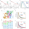
Fig. 1. smFRET analysis of solution conformations of the ileS T-box riboswitch.
a Four classes of T-box riboswitches with different structural features. IDTM: interdigitated double T-loop motif. T-box-tRNA contacts are indicated by red sticks. b Sequence and secondary structure of the two-piece Nocardia farcinica ileS T-box riboswitch construct used in smFRET analyses. Fluorophore labeling positions are indicated. c Co-crystal structure of the N. farcinica ileS T-box riboswitch in complex with tRNAIle (PDB: 6UFM). d Rotated, detailed view of the stems I–II docking interface showing the ribose zipper interactions. tRNA is omitted for clarity. Hydrogen bonds are indicated by dashed lines. e Isothermal titration calorimetry (ITC) measurement of tRNAIle binding to the WT two-piece T-box riboswitch construct. f Schematic of free-diffusion confocal smFRET analysis. gi FRET efficiency histograms of g WT T-box RNA with 10 mM Mg2+ in the absence or presence of 10 nM, 100 nM, or 1 μM tRNA, h WT T-box RNA with 1 mM Mg2+ in the absence or presence of 10 μM tRNA, and i G70U T-box RNA with 10 mM Mg2+ in the absence or presence of 1 μM tRNA. The histograms were constructed using bins (2 ms bin time) containing 30 photons or more. The peak at E ~ 0.1 (light gray) in the histograms results from the molecules lacking the acceptor. FRET efficiencies were corrected for background. Source data are provided as a Source Data file.
Fig. 2
Fig. 2. SAXS analyses and 2-aminopurine (2AP) lifetime probing of the T-box RNA.
a, b Overlay of back-calculated SAXS scattering curves (red dotted lines) computed from the cocrystal structure of the N. farcinica ileS T-box riboswitch in complex with tRNAIle (a) or the T-box portion extracted from the complex structure (b) using CRYSOL, with experimental SAXS profiles (black dotted lines). Residuals and χ2 of the fits are indicated. c Pair-wise distance distribution function (PDDF) plots for the tRNA (green), apo T-box (blue), and T-box-tRNA complex (black). d Location of the 2AP probes (A16, orange; A19, red) in the co-crystal structure at the T-box Specifier-tRNA anticodon interface. e, f Time-resolved fluorescence decay time traces for 2AP at positions A16 (e) or A19 (f). Amplitude-weighted averaged lifetimes τavg are indicated. IRF: instrument response function. g Individual lifetimes and relative amplitudes derived from (e, f). The amplitudes for the short, intermediate, and long lifetimes are shown in light blue, green, and red, respectively. Source data are provided as a Source Data file.
Fig. 3
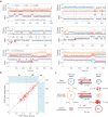
Fig. 3. Two-color immobilization smFRET experiment of the T-box riboswitch.
a Schematic of smFRET experiment of immobilized T-box RNA in the presence or absence of tRNA. b, d Representative binned (50 ms bin time) donor (AF488, blue) and acceptor (AF 594, orange) fluorescence trajectories (upper panel) and FRET efficiency trajectories (lower panel) for immobilized T-boxes in the absence (b) and presence (d) of tRNA, respectively. c, e Upper panels: FRET efficiency histograms of three different states in the absence (c) and presence (e) of 50 nM tRNA. FRET efficiency values were corrected for background, donor leak into the acceptor channel, γ-factor, and direct acceptor excitation (see Methods). Lower panels: 2-dimensional (2D) FRET efficiency-donor lifetime plots in the absence (c) and presence (e) of 50 nM tRNA. The distributions were clustered into three states using Gaussian mixture models. The deviation from the diagonal line indicates rapid fluctuations of donor-acceptor distances (i.e., conformational flexibility) in each state. The gray solid curve shows the dependence of the lifetime on the FRET efficiency for a flexible random polymer (Gaussian chain) for comparison. f Transition density plot between the three different states of WT T-box in the presence of 50 nM tRNA. Source data are provided as a Source Data file.
Fig. 4
Fig. 4. Three-color smFRET spectroscopy of tRNA ASL binding to the T-box RNA.
a Representative binned (50 ms bin time) donor (AF488, blue), acceptor 1 (AF594, orange), and acceptor 2 (CF680R, red) fluorescence (upper) and FRET efficiency trajectories (lower). The FRET efficiency is defined as the fraction of total acceptor photons (acceptor 1 and acceptor 2), which are corrected for background, leaks, γ-factors, and direct excitation of acceptors (see Methods). Due to incomplete acceptor 2 labeling on the ASL, the bound state can appear as both 2- (binding to unlabeled ASL) and 3-color (binding to labeled ASL) segments as indicated by green and red asterisks above the FRET trajectories, respectively. After dissociation of ASL, in most cases, T-box molecules come back to the unbound state with the FRET efficiency values similar to those before binding (i.e., same unbound state) unless infrequent changes occur either in the unbound state (red arrows) or in the bound state (between the two blue arrows), indicating the presence of long-lasting memory. b Transition density map analysis. Correlation of the efficiency (i.e., fraction of total acceptor photons) values of the unbound T-box before ASL binding and after its dissociation (i.e., E values of two unbound states separated by a bound state). The distribution appears along the diagonal line for almost all the cases of the open (U1) state, and most cases of semi-open (U2) states, indicating the memory of the unbound conformation persists through the bound state. Conversely, the memory of the closed state (U3) is frequently lost in the bound state (transitions in the blue-shaded off-diagonal regions). c Kinetic model for tRNA ASL binding to the T-box RNA showing rates of interconversion (as lifetimes) between the different states (three bound states B1–B3 and three unbound states U1–U3) in the presence of 20 nM tRNA-ASL (see Supplementary Fig. 8 for the complete kinetic model accounting for incomplete labeling of acceptor 2). The area of the circle is proportional to the population of each state, and the thickness of the arrows is proportional to the rate. *binding rates with 20 nM ASL. Source data are provided as a Source Data file.
Fig. 5
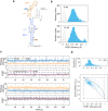
Fig. 5. smFRET spectroscopy of the ΔΨ T-box RNA.
a Sequence and secondary structure of the ΔΨ T-box RNA (nts 1–77, pseudoknot deletion). The location of the inserted uridine linker between stems I and II is indicated in red. b FRET efficiency histograms from free-diffusion experiments of ΔΨ T-box RNA in the absence (top) and presence (bottom) of 10 μM tRNA. FRET efficiency values were corrected for background. c Representative binned fluorescence (upper) and FRET efficiency (lower) trajectories of immobilized ΔΨ T-box RNA in the absence of tRNA. d Upper panel: FRET efficiency histogram of immobilized ΔΨ T-box RNA in the absence of tRNA, derived from (c). FRET efficiency values were corrected for background, donor leak into the acceptor channel, γ-factor, and direct acceptor excitation (see Methods). Lower panel: 2D FRET efficiency-donor lifetime plot of immobilized ΔΨ T-box RNA without tRNA. The on-diagonal distribution indicates a lack of flexibility in ΔΨ T-box. Source data are provided as a Source Data file.
Fig. 6
Fig. 6. Solution conformation of ΔΨ T-box RNA.
a Cartoon schematic (left) and structural models (right) of the ΔΨ T-box RNA rigid-body docked into the SAXS-reconstructed envelope generated by DAMMIF (gray), in stems I-II docked (upper panel) and stacked (lower panel) conformations. b Overlay of back-calculated SAXS scattering curves (red lines) computed using CRYSOL from the stems I-II docked structures extracted from the cocrystal structure (upper panel) or from the proposed stems I-II stacked structural model (lower panel) with experimental scattering profiles in solution (black lines). Residuals and χ2 of the fits are indicated. c PDDF plots for the apo T-box (gray) and ΔΨ T-box (black). d Effects of inserting a flexible polyuridine linker of different lengths between stems I and II of ΔΨ T-box on tRNA binding affinity. The values are mean ± S.D. from N biologically independent replicates. N = 3 for linker lengths 1 and 10; N = 2 for linker lengths 2–5. * value for zero U-linker length from ref. for comparison. Source data are provided as a Source Data file.
Fig. 7
Fig. 7. Mechanistic model of tRNA binding to a dynamic T-box mRNA ensemble.
Most initial T-box transcripts comprise three discrete, stable helical elements: stems I (orange), II (blue), and IIA/B pseudoknot (cyan, Ψ) linked by short or zero-length linkers. Due to strong thermodynamic motivations for helical termini to coaxially stack, stem I and the pseudoknot compete for end-to-end stacking with stem II. We posit that this competition is chiefly responsible for the creation of a dynamic mRNA conformational ensemble. Among the open, semi-open, and closed conformers, tRNA mostly engages the two open T-box conformers due to reduced steric hindrance and their larger populations. tRNA engagement triggers rapid stems I–II closure and docking, which traps the tRNA. In the absence of a stably folded pseudoknot, stems I and II stack instead, leading to a static conformation unable to retain the tRNA. Therefore, the polymorphic three-helix module of T-box mRNA is dynamically poised to rapidly recruit and stably retain its tRNA ligand, which in turn chaperones the folding of its multi-domain T-box RNA partner.

Similar articles

Cited by

References

    1. Kreuzer, K. D. & Henkin, T. M. The T-Box riboswitch: tRNA as an Effector to Modulate Gene Regulation. Microbiol. Spectr.10.1128/microbiolspec.RWR-0028-2018 (2018). - PMC - PubMed
    1. Suddala KC, Zhang J. An evolving tale of two interacting RNAs-themes and variations of the T-box riboswitch mechanism. IUBMB Life. 2019;71:1167–1180. doi: 10.1002/iub.2098. - DOI - PMC - PubMed
    1. Grundy FJ, Henkin TM. tRNA as a positive regulator of transcription antitermination in B. subtilis. Cell. 1993;74:475–482. doi: 10.1016/0092-8674(93)80049-K. - DOI - PubMed
    1. Gutierrez-Preciado A, Henkin TM, Grundy FJ, Yanofsky C, Merino E. Biochemical features and functional implications of the RNA-based T-box regulatory mechanism. Microbiol. Mol. Biol. Rev. 2009;73:36–61. doi: 10.1128/MMBR.00026-08. - DOI - PMC - PubMed
    1. Vitreschak AG, Mironov AA, Lyubetsky VA, Gelfand MS. Comparative genomic analysis of T-box regulatory systems in bacteria. RNA. 2008;14:717–735. doi: 10.1261/rna.819308. - DOI - PMC - PubMed

Publication types