. 2023 Aug 25;26(10):107709.
doi: 10.1016/j.isci.2023.107709. eCollection 2023 Oct 20.

Parasympathetic and sympathetic axons are bundled in the cardiac ventricles and undergo physiological reinnervation during heart regeneration

Affiliations

Parasympathetic and sympathetic axons are bundled in the cardiac ventricles and undergo physiological reinnervation during heart regeneration

Rebecca J Salamon et al. iScience. .

Abstract

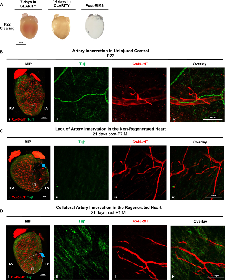
Sympathetic innervation influences homeostasis, repair, and pathology in the cardiac ventricles; in contrast, parasympathetic innervation is considered to have minimal contribution and influence in the ventricles. Here, we use genetic models, whole-mount imaging, and three-dimensional modeling to define cardiac nerve architecture during development, disease, and regeneration. Our approach reveals that parasympathetic nerves extensively innervate the cardiac ventricles. Furthermore, we identify that parasympathetic and sympathetic axons develop synchronously and are bundled throughout the ventricles. We further investigate cardiac nerve remodeling in the regenerative neonatal and the non-regenerative postnatal mouse heart. Our results show that the regenerating myocardium undergoes a unique process of physiological reinnervation, where proper nerve distribution and architecture is reestablished, in stark contrast to the non-regenerating heart. Mechanistically, we demonstrate that physiological reinnervation during regeneration is dependent on collateral artery formation. Our results reveal clinically significant insights into cardiac nerve plasticity which can identify new therapies for cardiac disease.

Keywords: Cell biology; Molecular biology; Neuroscience.


Conflict of interest statement

The authors declare no competing interests.

Figures

None
Graphical abstract
Figure 1
Figure 1
Parasympathetic and sympathetic axons are bundled and synchronous in development Parasympathetic reporter hearts (ChATCre;Rosa26tdTomato) were immunostained for sympathetic nerves with tyrosine hydroxylase (TH), imaged with confocal microscopy, and reconstructed with Imaris. (A) At postnatal day 7 (P7), (Ai) whole-heart max intensity projection (MIP) of the posterior mature nerve architecture shows close association between parasympathetic and sympathetic axons, with (Aii) region of interest (ROI) demonstrating large and small nerve fibers closely aligned. (Aiii) ROI inset (white box in Aii) and (Aiv) their cross-section view (white dashed line in Aiii overlay) of axon bundles shows distinct parasympathetic and sympathetic axons (n = 7). (B) At embryonic day 16.5 (E16.5), (Bi) both parasympathetic and sympathetic axons extend together in the posterior wall, with (Bii) high-magnification and (Biii) a cross-section view (white dashed line in Bii overlay) demonstrating close localization and bundling of nerve subtypes (n = 6). (C and D) 3D modeling and analysis shows increased branching level of parasympathetic and sympathetic nerves (PSN, SN) during embryonic and postnatal development (n = 5–7). (E) Density analysis of the P7 heart shows axon bundles of primary and secondary axons are at higher density than tertiary and higher branched axons. Right and Left Ventricles (RV, LV) are indicated. Scale bars shown at 1 mm for low magnification, 100 μm for high magnification, and 10 μm for insets. Groups compared by an ordinary two-way ANOVA, with uncorrected Fisher’s LSD, with a single pooled variance. Significance shown as n.s. (p > 0.05), ∗ (P 0.05). ∗∗(P 0.01), ∗∗∗(P 0.001), ∗∗∗∗ (P 0.0001). Data are represented as mean ± SEM. Sample size (n) represents the number of animals. See also Figures S1–S3.
Figure 2
Figure 2
Neurovascular association shows cardiac axons align with the coronary arteries Embryonic day 17.5 (E17.5) hearts of Cx40CreER;Rosa26tdTomato coronary artery reporter mice were immunostained with the pan-neuronal marker Tuj1 and the endothelial cell marker endomucin (EMCN), followed by whole-mount confocal imaging, and 3D reconstruction and modeling. (A) The neurovascular patterning is shown as (Ai) a whole-heart max intensity projection (MIP) of the anterior embryonic heart and (Aii) a high-magnification in region of interest (ROI) (Aiii) 3D Imaris reconstruction of the whole embryonic heart, and (Aiv) a reconstruction of the ROI (n = 6). (B) Neurovascular depth analysis shows that the anterior nerves are at similar depths to the coronary arteries (n = 3). (C) Quantification of percentage of nerves associated within 100 μm of coronary veins (blue), arteries (red) both veins and arteries (gray) or neither vessel type (green) are shown, with an increased nerve artery association in the anterior wall (n = 6). Right and Left Ventricles (RV, LV) are indicated. Scale bars shown at 1 mm for low magnification and 100 μm for high magnification. Groups compared by an ordinary two-way ANOVA, with uncorrected Fisher’s LSD, with a single pooled variance. Significance shown as n.s. (p > 0.05), ∗ (P 0.05). ∗∗(P 0.01), ∗∗∗(P 0.001), ∗∗∗∗ (P 0.0001). Data are represented as mean ± SEM. Sample size (n) represents the number of animals. See also Figure S4 and Video S1.
Figure 3
Figure 3
Mature 3D neurovascular architecture P7 hearts of Cx40CreER;Rosa26tdTomato coronary artery reporter mice were immunostained with the pan-neuronal marker Tuj1 and the endothelial cell marker endomucin (EMCN), followed by whole-mount confocal imaging, and 3D reconstruction. (A) The posterior wall distribution of the mature cardiac nerves is shown as a whole-heart max intensity projection (MIP) of patterning of (Ai) posterior nerve patterning and (Aii) nerve-vein association. (Aiii-iv) Region of interest (ROI) shows the nerves align without innervating the major left, medial, or right coronary veins (LCV, MCV, RCV), with (Av) a representative magnified inset demonstrating lack of vein innervation (white box in Aiv) (n = 4). (B) The anterior nerve architecture similarly is shown as MIP of the (Bi) anterior nerve patterning and (Bii) nerve-artery association. (Biii-iv) Nerves align and directly innervate the right and left coronary arteries (RCA, LCA), with (Bv) a representative magnified z-plane inset (white box in Biv) and (Bvi) a graphical representation demonstrating direct artery innervation (n = 5). (C) Imaris 3D reconstruction highlights the neurovascular architecture of the posterior and anterior heart (n = 3). (D) Quantification of percent of nerves associated within 100 μm of coronary veins (blue), arteries (red) both veins and arteries (gray) or neither vessel type (green) are shown, with an increased nerve artery association in the anterior wall (n = 4). Scale bars shown at 1 mm for low magnification, 100 μm for high magnification, and 20 μm for insets. Right and Left Ventricles (RV, LV) are indicated. Groups compared by an ordinary two-way ANOVA, with uncorrected Fisher’s LSD, with a single pooled variance. Significance shown as n.s. (p > 0.05), ∗ (P 0.05). ∗∗(P 0.01), ∗∗∗(P 0.001), ∗∗∗∗ (P 0.0001). Data are represented as mean ± SEM. Sample size (n) represents the number of animals.
Figure 4
Figure 4
Cardiac axons show unique plasticity during neonatal heart regeneration Myocardial Infarction (MI) was performed on WT mice at regenerative (P1) and non-regenerative (P7) timepoints. Nerve remodeling was compared to uninjured, age-matched control hearts. (A) P22 uninjured hearts show control innervation in regions similar to the remote zone (RZ, pink), border zone (BZ, green), and infarct zone (IZ, navy blue). (B–D) Regenerating hearts were collected following P1 MI at 7, 14, and 21 days post MI (DPMI). (B) At 7DPMI, regenerative hearts were hyperinnervated in the RZ and denervated in the BZ and IZ (n = 5), in comparison to P8 uninjured hearts (n = 5). (C) At 14DPMI, regenerative hearts were hyperinnervated in the RZ, with restored innervation in the BZ and decreased innervation in the IZ (n = 4), in comparison to P15 uninjured hearts (n = 5). (D) At 21DPMI, regenerated hearts show restored levels of innervation in the RZ, BZ, and IZ (n = 5), in comparison to P22 uninjured hearts (n = 5). (E) Non-regenerative hearts were collected following P7 MI at 21DPMI. Hearts showed no significant difference in RZ innervation, with heterogeneous innervation in the BZ and denervation in the IZ, both indicators of nerve pathology (n = 4). In whole-heart images, dashed borders outline the RZ (pink), BZ (green), and IZ (navy blue), and the light blue knot represents the suture site. Right and Left Ventricles (RV, LV) are indicated. Scale bars shown at 1 mm for low magnification and 100 μm for high magnification. Groups compared using ordinary one-way ANOVA with Tukey post hoc test. Significance shown as n.s. (p > 0.05), ∗ (P 0.05). ∗∗(P 0.01), ∗∗∗(P 0.001), ∗∗∗∗ (P 0.0001). Data are represented as mean ± SEM. Sample size (n) represents the number of animals.
Figure 5
Figure 5
Parasympathetic and sympathetic nerves precisely reinnervate the regenerating heart Myocardial Infarction (MI) was performed on ChATCre;Rosa26tdTomato parasympathetic nerve reporter mice at regenerative (P1) and non-regenerative (P7) timepoints. Hearts were collected at 21 days post MI, and immunostained for tyrosine hydroxylase (TH) and imaged with confocal microscopy. (A) Uninjured hearts in adult (P22) mice show parasympathetic and sympathetic nerve bundling in (Ai) whole-heart max intensity projection (MIP) of low magnification and (Aii) high magnification views of the ventricles (n = 6). (B) Non-regenerating hearts show pathological remodeling of the nerves at 21 days post P7 MI. (Bii) The border zone (BZ) shows sympathetic axon sprouting (indicated by white arrows), independent of parasympathetic axons. (Biii) The infarct zone (IZ) is denervated (n = 6). (C) The regenerated heart shows reestablished parasympathetic and sympathetic axon bundling at 21 days post P1 MI. (Cii) The BZ shows dense nerve bundles of both nerve subtypes. (Ciii) The IZ also shows reinnervation of parasympathetic and sympathetic axons (n = 5). In whole-heart MIP images, white dashed border indicates the IZ and the light blue knot represents the suture site. Right and Left Ventricles (RV, LV) are indicated. Scale bars shown at 1 mm for low magnification and 100 μm for high magnification. Sample size (n) represents the number of animals used.
Figure 6
Figure 6
Nerve-artery association is reestablished in the regenerated heart MI was performed in Cx40CreER;Rosa26tdTomato coronary artery reporter mice at regenerative (P1) or non-regenerative (P7) timepoints. (A) Hearts were collected at 21 days post MI and underwent tissue clearing and nerve immunostaining with Tuj1. (B) Hearts were compared to uninjured P22 hearts, which showed (Bii) nerves, (Biii) arteries, and (Biv) artery innervation (n = 4). (C) Non-regenerative hearts at 21 days post P7 MI showed (Cii) denervation in the infarct zone (IZ), (Ciii-iv) including areas with vascularization (n = 6). (D) Regenerative hearts at 21 days post P1 MI showed (Dii) innervation in the IZ, (Diii-iv) including reinnervation of arteries in the IZ (n = 6). In whole-heart MIP images, dashed white lines indicate the IZ and the light blue knot represents the suture site. Right and Left Ventricles (RV, LV) are indicated. Scale bars shown at 1 mm for low magnification and 100 μm for high magnification. Sample size (n) represents the number of animals used. See also Figure S5.
Figure 7
Figure 7
Reinnervation of the regenerating heart is dependent on collateral artery formation MI was performed on Cx40CreER;Rosa26tdTomato mice during the regenerative window (P2) and hearts were collected at 21-days post MI. (A and B) We identify that the arteries in the infarct zone are reinnervated by 21-days post MI. To determine whether reinnervation during regeneration is dependent on collateral artery formation, we used the inducible Cx40CreER;Cxcr4fl/fl mouse to inhibit migration of arterial cells post MI. At 21 days post MI, Tuj1 immunostaining showed (B) innervation in the infarct zone (IZ) of wild type (WT) regenerated hearts (n = 8), as expected. (C) In the Cx40CreER;Cxcr4fl/fl mice, the IZ remained denervated, shown by (Ci) whole-heart max intensity projection (MIP) and (Cii) high-magnification of the IZ (n = 4). In whole-heart image, navy blue dashed border indicates the IZ and the light blue knot represents the suture site. Right and Left Ventricles (RV, LV) are indicated. Scale bars shown at 1 mm for low magnification and 100 μm for high magnification. Significance shown as n.s. (p > 0.05), ∗ (P 0.05). ∗∗(P 0.01), ∗∗∗(P 0.001), ∗∗∗∗ (P 0.0001). Data are represented as mean ± SEM. Sample size (n) represents the number of animals.

Update of

References

    1. Hasan W. Autonomic cardiac innervation: development and adult plasticity. Organogenesis. 2013;9:176–193. doi: 10.4161/org.24892. - DOI - PMC - PubMed
    1. Randall W.C. Oxford University Press; 1984. Nervous Control of Cardiovascular Function.
    1. Randall W.C. Williams and Wilkins Company; 1965. Nervous Control of the Heart.
    1. Achanta S., Gorky J., Leung C., Moss A., Robbins S., Eisenman L., Chen J., Tappan S., Heal M., Farahani N., et al. A Comprehensive Integrated Anatomical and Molecular Atlas of Rat Intrinsic Cardiac Nervous System. iScience. 2020;23 doi: 10.1016/j.isci.2020.101140. - DOI - PMC - PubMed
    1. Hanna P., Dacey M.J., Brennan J., Moss A., Robbins S., Achanta S., Biscola N.P., Swid M.A., Rajendran P.S., Mori S., et al. Innervation and Neuronal Control of the Mammalian Sinoatrial Node a Comprehensive Atlas. Circ. Res. 2021;128:1279–1296. doi: 10.1161/CIRCRESAHA.120.318458. - DOI - PMC - PubMed

LinkOut - more resources